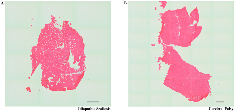
Figure 3.
Representative hematoxylin and eosin (H&E) stained muscle tissue. H&Es from a patient with idiopathic scoliosis (panel (A)) and cerebral palsy (panel (B)) correspond to tissues from which images represented in Figure 4 were obtained. Whole tiled images were captured at 10× magnification on an Evos microscope (scale bar in each photo represents 500 µM.) These images show the general morphology of the muscle samples used in the study.

