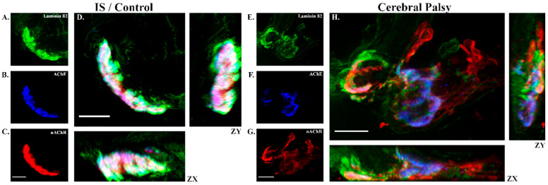
Figure 4.
Representative neuromuscular junctions from control and cerebral palsy. Skeletal muscle cryosections were immunofluorescently labeled to illuminate the co-localization of laminin β2 (green; panels (A,E)), AChE (blue; panels (B,F)), and nAChR (red; panels (C,G)). Co-staining displays as white or lavender tones (D,H) when the color channels are highly co-localized. On average, NMJs from participants with CP (e.g., right panel) exhibited significantly disrupted NMJ organization as compared to controls (left panel; n = 200 total NMJs, overall). Disorganization among CP NMJs was evident in the ZY-ZX planes as well. Scale bar represents 10 µM.

