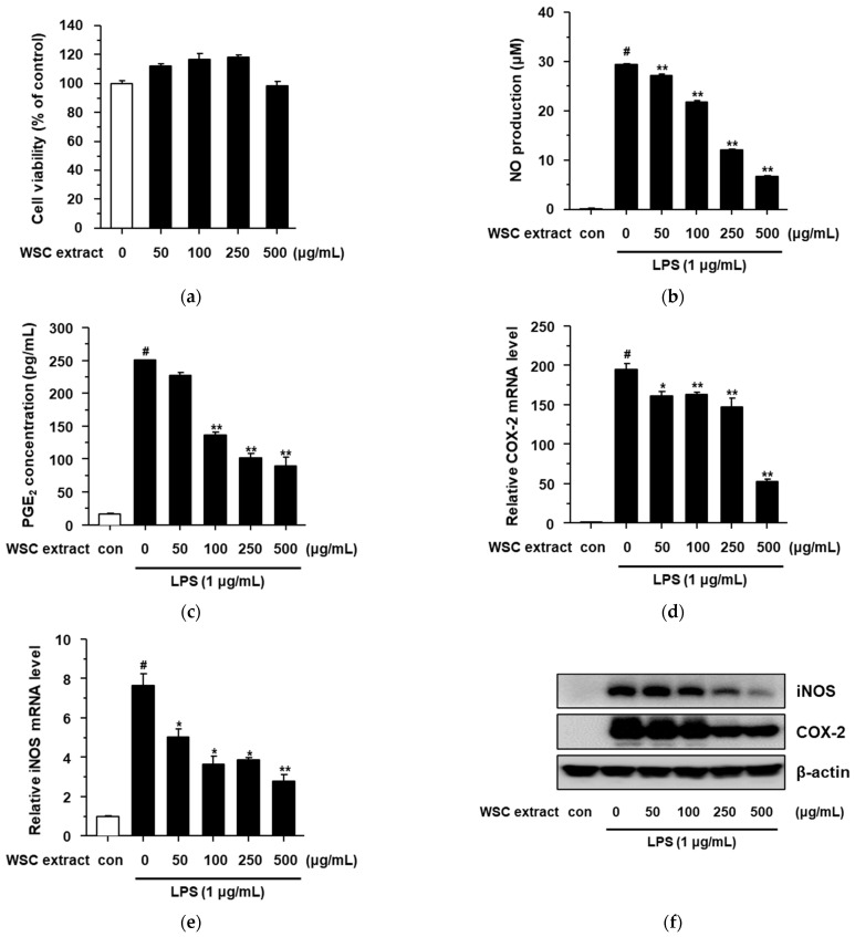
Figure 3.
Anti-inflammatory effects of the WSC extract on LPS-stimulated RAW264.7 cells: (a) Cytotoxicity of the WSC extract was measured via dimethylthiazol-diphenyltetrazolium bromide assay; (b) Griess reaction assay was performed to determine NO production; (c) PGE2 concentration was determined using an ELISA kit; (d,e) iNOS and COX-2 mRNA levels were analyzed by quantitative real-time PCR; (f) iNOS and COX-2 protein levels were determined by western blotting assay. Values are expressed as the mean ± standard deviation of three independent experiments; #, p < 0.005 versus control; *, p < 0.05; **, p < 0.005 versus LPS alone.

