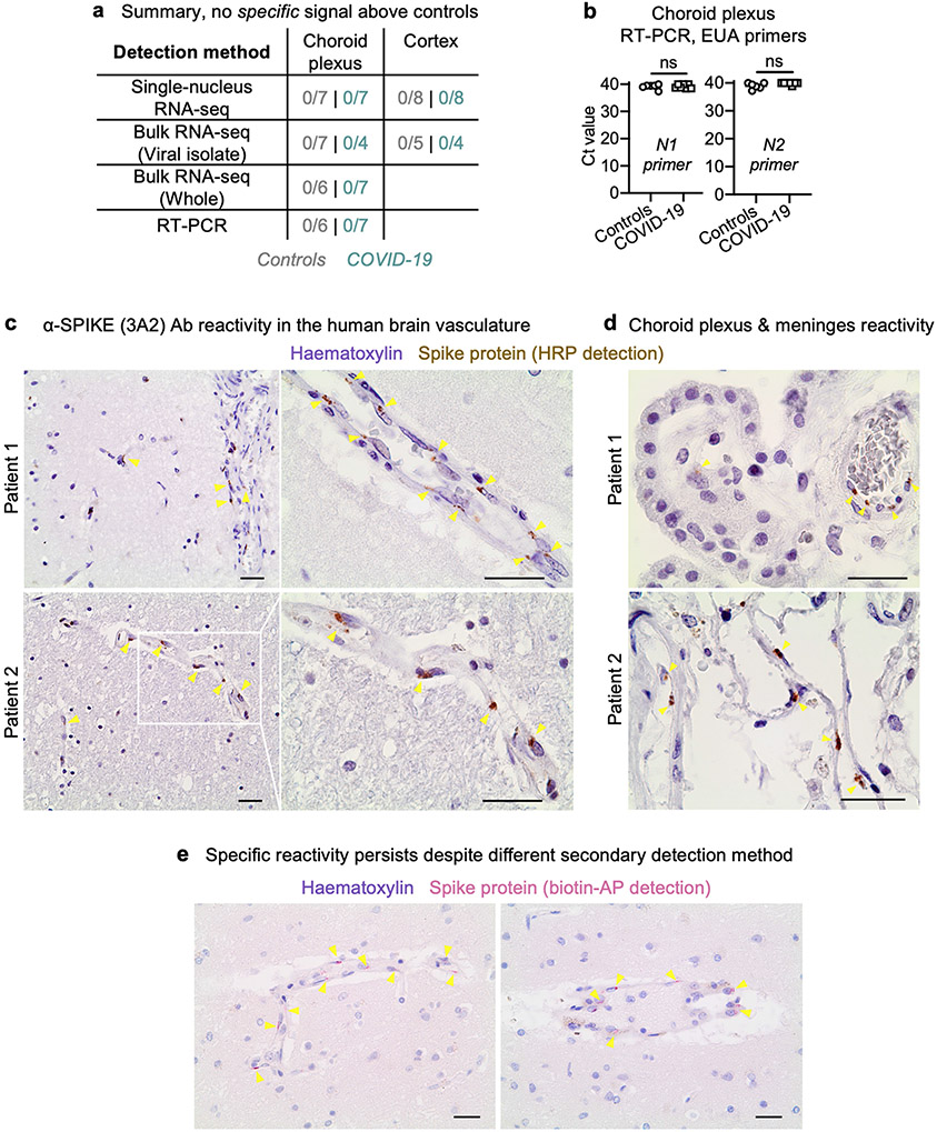
Extended Data Fig. 9 ∣. No conclusive detection of SARS-CoV-2 neuroinvasion.
a, Summary of RNA-based assays to detect SARS-CoV-2 in the human cortex and choroid plexus. Aside from the 3A2 antibody, no other anti-SARS-CoV-2 antibody detected viral protein antigen in the brain or choroid plexus. b, qPCR detection of the SARS-CoV-2 genes N1 and N2 via CDC Emergency Use Authorization primers on choroid plexus samples (n = 6 non-viral control, n = 7 COVID-19; two-sided Mann–Whitney t-test; mean ± s.e.m.). c, Aberrant anti-SARS-CoV-2 spike (3A2) antibody reactivity (brown) in the frontal medial cortex of two patients with COVID-19 in tissue immediately adjacent to that used for snRNA-seq. Haematoxylin counterstain (purple). Scale bar, 20 μm. d, As in c, but for the choroid plexus and meninges in two patients with COVID-19. Scale bar, 20 μm. e, As in c, but using a different secondary antibody detection method (biotin–alkaline phosphatase (red)), recapitulating the specific vascular-localized signal. Scale bar, 20 μm. Immunohistochemical stains are representative of at least two independent experiments.

