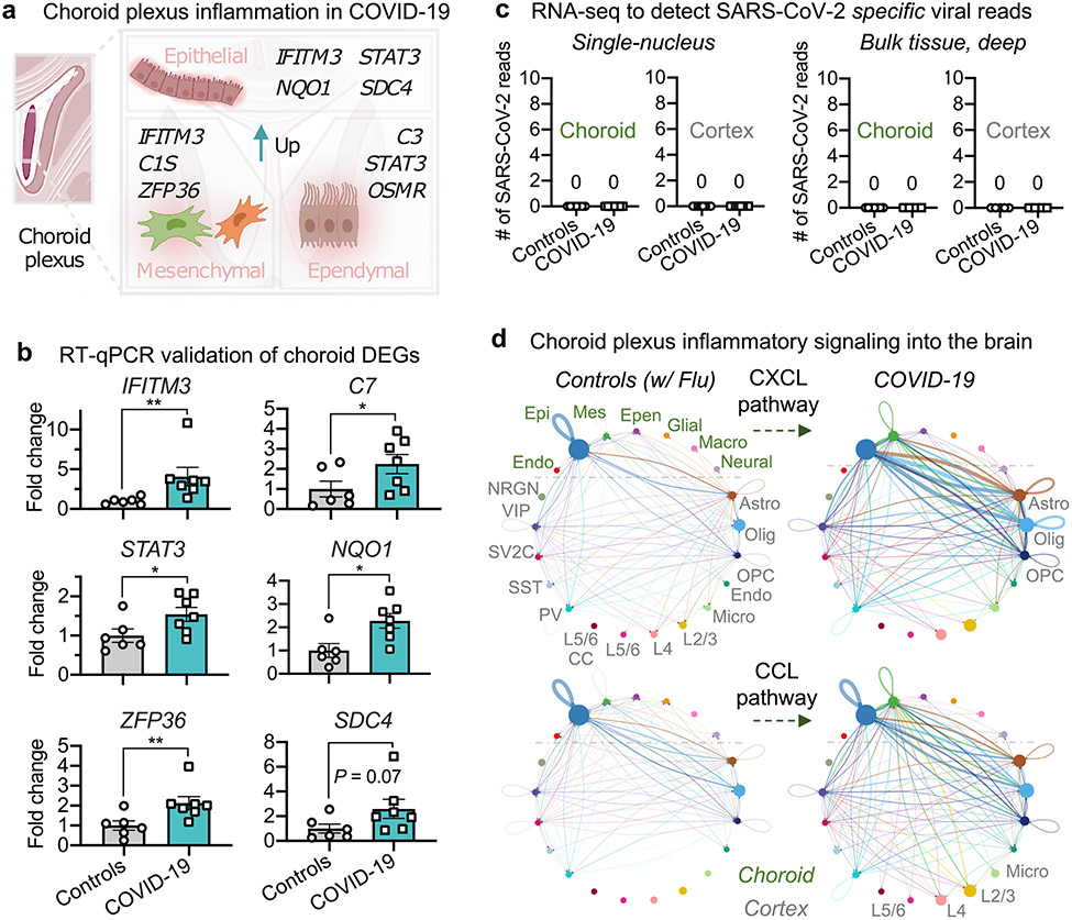
Fig. 2 ∣. Brain-barrier inflammation in patients with COVID-19 does not require direct replicative infection.
a, Examples of inflammation-related DEGs in the choroid plexus of patients with COVID-19 (n = 6 control individuals (without viral infection); n = 7 patients with COVID-19; MAST with default settings). DEGs defined as log-transformed fold change > 0.25 (absolute value) and adjusted P value < 0.05 (Bonferroni correction). b, Validation of predicted choroid plexus DEGs by RT–qPCR (n = 6 control individuals (without viral infection), n = 7 patients with COVID-19; two-sided Mann–Whitney t-test; mean ± s.e.m.). Genes chosen for validation are either immediately related to SARS-CoV-2 (IFITM3) or genes with log-transformed fold changes similar to those of IFITM3 (NQO1), to assess the robustness of snRNA-seq thresholds. P values P = 0.0023 (IFITM3), 0.0484 (C7), 0.0350 (STAT3), 0.0140 (NQO1), 0.0082 (ZFP36) and 0.0734 (SDC4). c, snRNA-seq (left) or bulk RNA-seq (right) of choroid plexus and cortex from control individuals or patients with COVID-19 (no reads). snRNA-seq, n = 7 control, n = 7 COVID-19 (choroid plexus); n = 7 control, n = 7 COVID-19 (cortex). Bulk RNA-seq (after viral RNA isolation): n = 7 control, n = 4 COVID-19 (choroid plexus); n = 5 control, n = 4 COVID-19 (cortex). d, Circle plot showing the number of statistically significant intercellular signalling interactions for the CXCL and CCL pathway family of molecules in control individuals compared to patients with COVID-19 (permutation test, CellChat32; n = 8 control individuals (including patients with influenza); n = 8 patients with COVID-19 (cortex); and n = 7 control individuals (including patients with influenza); n = 7 patients with COVID-19 (choroid plexus)). Each circle (colour) represents one cell type; edges connecting circles represent significant intercellular signalling inferred between those cell types. Circles and edges are normalized to the number of cells for a given cell type and inferred strength of signalling, respectively. Cell types labelled on the right correspond to signalling pathways increased in COVID-19. Endo., endothelial; epen., ependymal; epi., epithelial; mes., mesenchymal.

