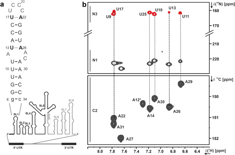
Fig. 1.
a Secondary structure of 5_SL1 and its genomic position within the 5'-UTR of the SARS-CoV-2 genome. b Detection of the W–C base-pairs U13-A26 and U17-A22 in the lrHNN-COSY experiment (Table 1, XIII.). Adenosine C2H2 resonances (lower spectrum, 1H,13C-HSQC) were used to assign the 2J-N1H2 diagonal peaks and the corresponding uridine N3 cross peaks. Note that the A12 N1H2 resonance is broadened beyond detection. The U13-A22 and U17-A22 correlations are shown in black, the other base-pairs in grey in panel a

