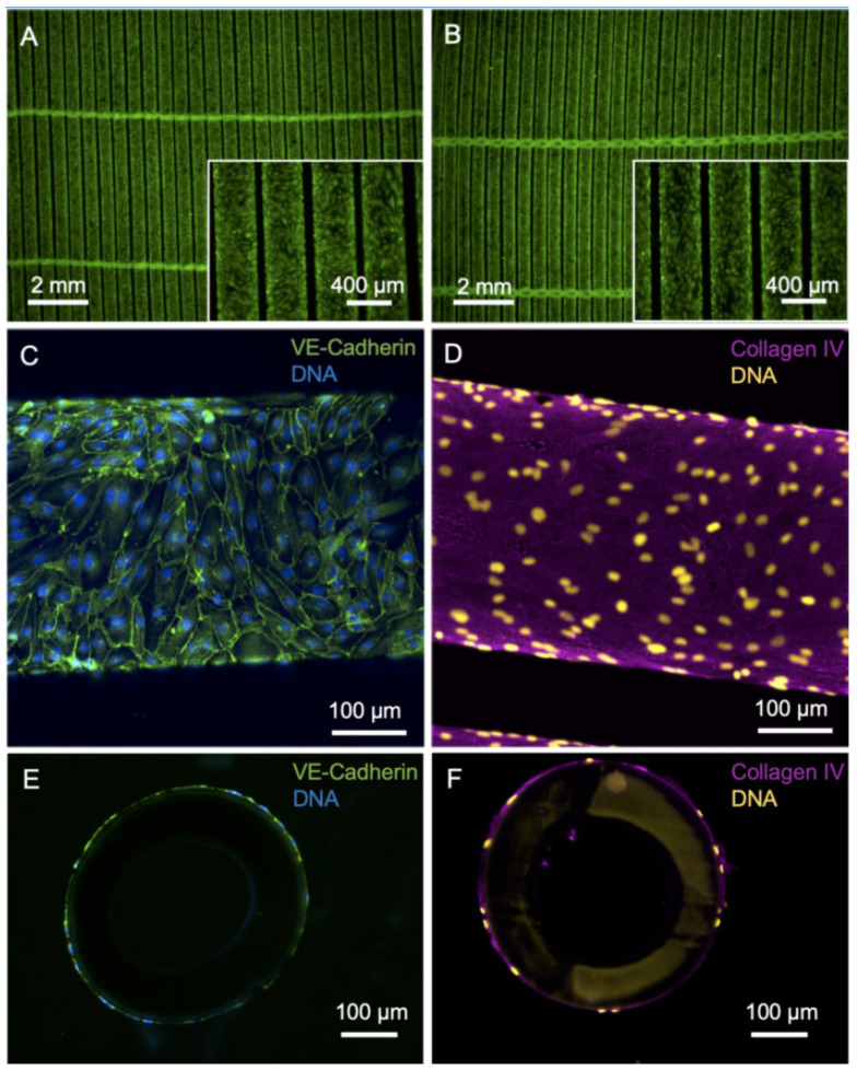
Figure 1.
Imaging of HFM after endothelialization with iPSC-EC1. (A,B) Viable calcein-positive iPSC-ECs (green) seeded on fibronectin-coated HFM form a confluent monolayer around the individual fibers of the HFM. A: Top view, B: bottom view. (C) iPSC-ECs are interconnected via VE-cadherin (green). (D) Basal-lamina-like matrix envelops iPSC-EC-seeded hollow fibers, indicated by de novo synthesized collagen type-IV (magenta). (E,F) Nuclei counterstained with DAPI and presented in false colors (yellow).

