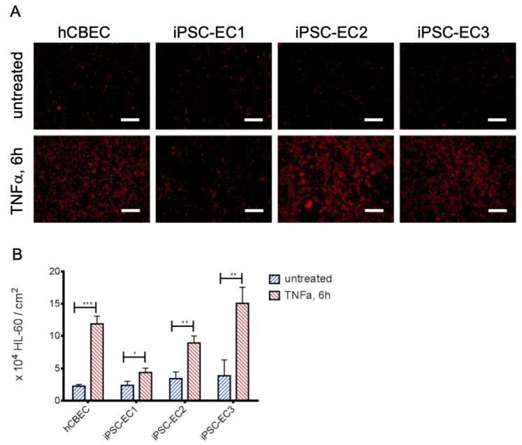
Figure 3.
iPSC-ECs on PMP membranes show different levels of leukocyte adhesion after TNFα treatment. (A) Exemplary images of cell tracker red-stained HL-60 leukocytes (red) on iPSC-EC endothelialized gas exchange membranes under standard condition and after treatment with TNFα for 6 h. Except for monolayers established from iPSC-EC1s, appreciably increased numbers of HL-60 cells adhered to EC-monolayers incubated with TNFα. Scalebar: 250 µm. (B) Quantification of adhered HL-60 leukocytes. Reported values are given as means ± standard deviation. Significant differences between untreated and TNFα-stimulated groups were marked with * at p < 0.05, ** when p < 0.01 and with *** when p < 0.001.

