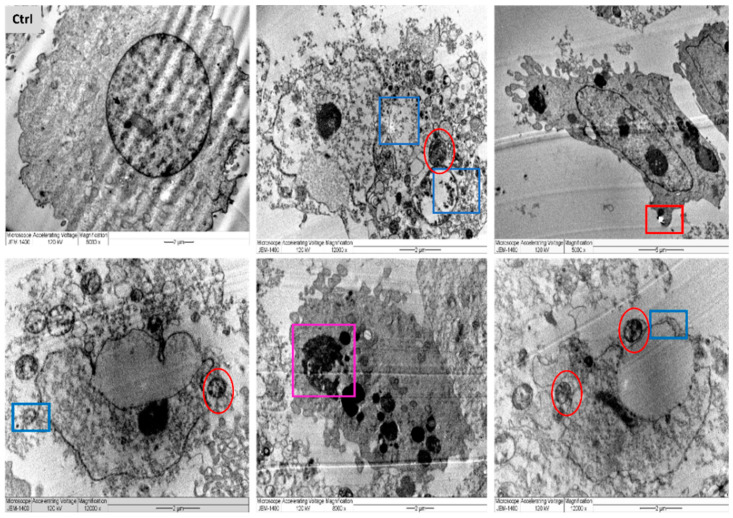
Figure 14.
TEM images of MDA-MB-231 cell lines, where untreated cell (Ctrl) exhibiting rounded shape cell with complete organelle and nucleus. Image magnification ×6.000; bar 2 μm. Other images at varied magnification show the ultrastructural alteration in cell treated with R-AgNPs displaying early apoptosis characteristics such as: chromatin condensation and over whole cell shrinkage as well as late apoptosis: lipid droplet (red square), peroxisomes (yellow square) and enlarged mitochondria (yellow circle), damaged mitochondria (red circle) and condensed nucleus (Purple square). Damaged cancerous cells are clearly observed with R-AgNPs located in the cytoplasm, outer cell and nucleus membranes (blue squares).

