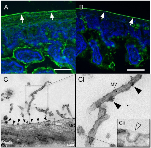
Figure 2.
Mesopolysaccharide (MPS) of murine small bowel MPS. The glucose/mannose group lectin Concanavalin ensiformis (ConA) [34] (A) and the N-acetylglucosamine group lectin Solanum tuberosum (STA) [35] (B) were used to demonstrate carbohydrates on the serosal surface (white arrows; bar = 50 µm). (C) Transmission electron microscopy (TEM) of the serosa demonstrating mesothelial cells (mc) and microvilli (mv). Higher resolution image (Ci) demonstrating findings consistent with MPS attached to the microvilli. Membrane vesicles and pits potentially involved in MPS hydration and maintenance are seen beneath the cell surface (Cii, inset).

