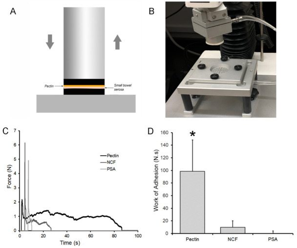
Figure 3.
Tensile strength adhesion testing ex vivo. (A,B) A custom fixture with a pectin mounted 30 mm flat surface probe was used to engage the 1 mm thick serosal specimen mounted on the fixture platform. After 5N compression for 30 s development time, the probe was withdrawn with 500 pps force and distance recordings. (C) Due to the extensibility of the subserosal tissue, the pectin-serosal adhesion resulted in a protracted adhesion curve (black line). In contrast, the nanocellulose fiber (NCF) film (dark gray) and pressure sensitive adhesive (PSA) (light gray) debonded shortly after the onset of withdrawal. (D) Replicate samples demonstrated that the work of adhesion, representing the area under the force-distance curve, was significantly greater in the pectin samples compared to NCF and PSA (*, p < 0.001).

