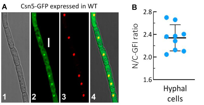
Figure 1.
Subcellular localisation of Csn5 in B. bassiana. (A) LSCM images (scale bar: 5 μm) for subcellular localisation of the Csn5–GFP fusion protein in the hyphal cells stained with DAPI nuclear dye (shown in red) after collection from 2-day-old SDBY culture. Panels 1, 2, 3, and 4 are bright, expressed, stained, and merged views of the same field. (B) Nuclear versus cytoplasmic green fluorescence intensity (N/C-GFI) ratios of the fusion protein measured from in the cells of nine hyphae.

