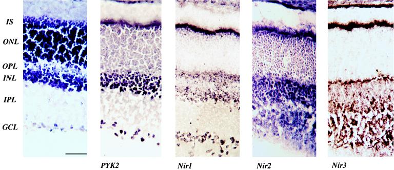
FIG. 6.
Distribution of the PYK2 and Nir proteins in the rat retina. (A) Hematoxylin staining of a vertical cryostat section of a rat retina showing different layers of the retina. (B) PYK2 immunoreactivity in the retina. Strong staining for PYK2 is detected throughout the inner nuclear layer (INL) and in the ganglion cell layer (GCL); specific staining is also observed in the outer nuclear layer (ONL). (C) Nir1 immunoreactivity in the retina. Strong staining is detected in the inner segment (IS), in the outer plexiform layer (OPL), in specific cells within the inner nuclear layer, and in the ganglion cell layer. (D) Nir2 immunostaining is seen in the inner segment, outer nuclear layer, and inner plexiform layer (IPL), and a moderate expression level is seen in the inner nuclear layer and the outer plexiform layer. (E) Strong immunostaining of Nir3 is detected in the inner segment and the inner and outer plexiform layers (scale bar, 50 μM).

