Figure 3.
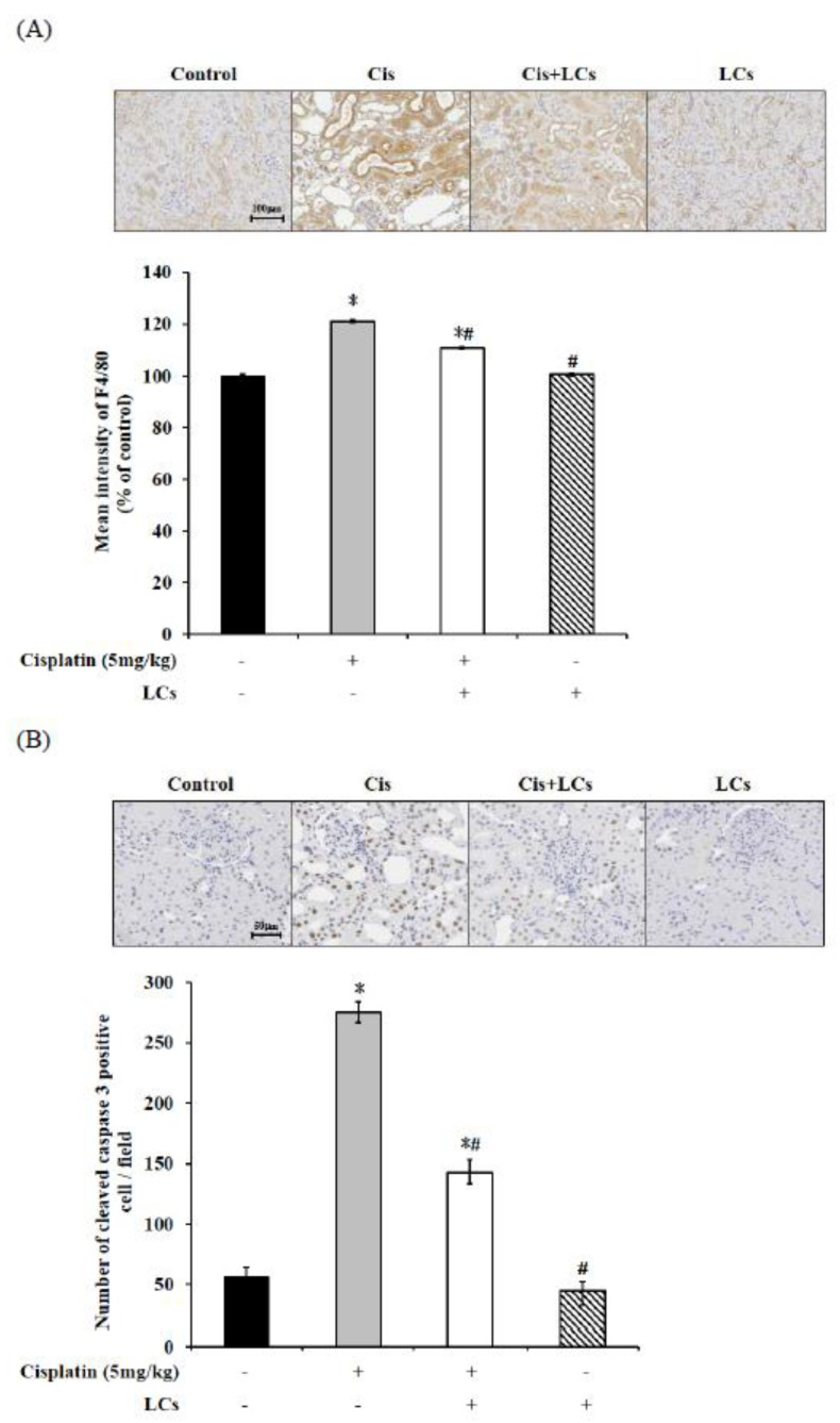
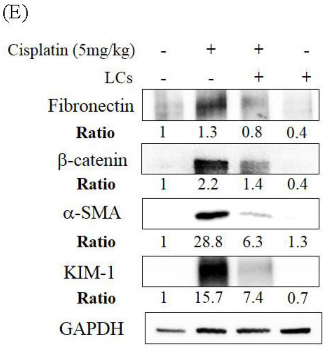
LCs supplementation alleviates renal fibrosis, necrosis, and macrophage infiltration in cisplatin-induced nephrotoxicity rats. (A) Immunohistochemistry shows F4/80+ macrophage infiltration and quantitative analysis. (B) Immunohistochemistry shows cleaved caspase-3-positive cells and quantitative analysis of tubular necrosis. (C) Hydroxyproline levels. (D) Immunohistochemistry shows collagen IV intensity and quantitative analysis of positive cells. (E) Western blot analysis shows renal fibronectin, β-catenin, α-SMA, and KIM-1 protein expression. Results are shown as mean ± SEM (n = 7 per group). * p < 0.05 compared with the control group; # p < 0.05 compared with the cisplatin group.


