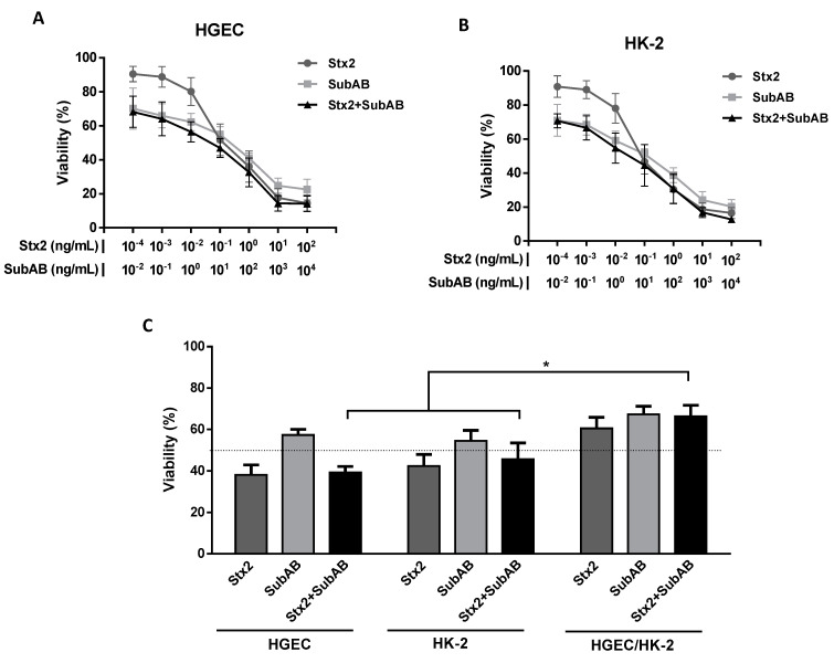
Figure 1.
Reduction of cell viability on HGEC and HK-2 cells by the co-incubation (Stx2 + SubAB). HGEC (A) and HK-2 (B) cells were incubated with different concentrations of Stx2 (1 × 10⁻⁴ to 1 × 10² ng/mL), SubAB (1 × 10⁻² to 1 × 10⁴ ng/mL) or with both toxins together (Stx2 + SubAB) in growth-arrested conditions for 72 h. Then, cell viability was analyzed by neutral red uptake. Absorbance in each well was read at 540 nm. One hundred percent represents cells incubated under identical conditions but without toxin treatment (Control). Results are expressed as means ± SEM, (n = 8). (C) HGEC and HK-2 monocultures and HGEC/HK-2 cocultures were exposed to the 50% cytotoxic dose (CD50) for Stx2 (0.1 ng/mL), SubAB (10 ng/mL) or Stx2 + SubAB (0.1 ng/mL + 10 ng/mL) for 72 h. Then, cell viability was analyzed by neutral red uptake. Results are expressed as the means ± SEM, (n = 9), * p < 0.05.

