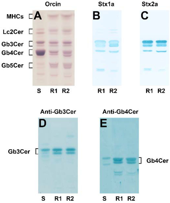
Figure 1.
Orcinol stain (A) and TLC overlay assays of the neutral GSL preparation obtained from pHRPTEpiCs using Stx1a (B) and Stx2a (C) as well as anti-Gb3Cer (D) and anti-Gb4Cer (E) antibody. The employed GSL quantities for TLC separation were equivalent to 5 × 10⁶ cells (A, orcinol stain), 6 × 10⁵ cells (B,C, Stx1a and Stx2a, respectively) and 2 × 10⁶ cells using the anti-Gb3Cer (D) and the anti-Gb4Cer antibody (E), respectively. S, 20 µg of a standard GSL mixture prepared from human erythrocytes (A), and 2 µg and 0.2 µg for the anti-Gb3Cer (D) and the anti-Gb4Cer (E) TLC immunodetection, respectively; R1, replicate 1; R2, replicate 2; MHCs, monohexosylceramides; Lc2Cer, lactosylceramide. Cells of the fifth passage were used for GSL isolation.

