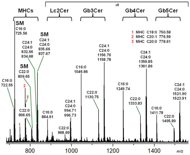
Figure 2.
MS1 spectrum of the sphingolipid preparation including neutral GSLs and sphingomyelins obtained from pHRPTEpiCs. The sphingolipids were isolated from cells of the 5th passage of biological replicate 2 (R2). The orcinol stain of the TLC-separated GSLs is shown in Figure 1A accompanied by the Stx1a, Stx2a, anti-Gb3Cer, and anti-Gb4Cer TLC overlay assays depicted in Figure 1B–E. Mono-, di-, tri-, tetra-, and pentahexosylceramides were identified as monohexosylceramides (MHCs), Lc2Cer, Gb3Cer, Gb4Cer, and Gb5Cer, respectively. The sphingolipids were detected as monosodiated [M+Na]+ species operating in the positive ion mode and are displayed in Table 1. Structural proofs were performed by CID experiments, and examples of MS2 spectra are given for Gb3Cer (d18:1, C22:0), Gb4Cer (d18:1, C16:0), and proposed Gb5Cer (d18:1, C22:0) in Figures S2–S4, respectively, in the Supplementary Materials.

