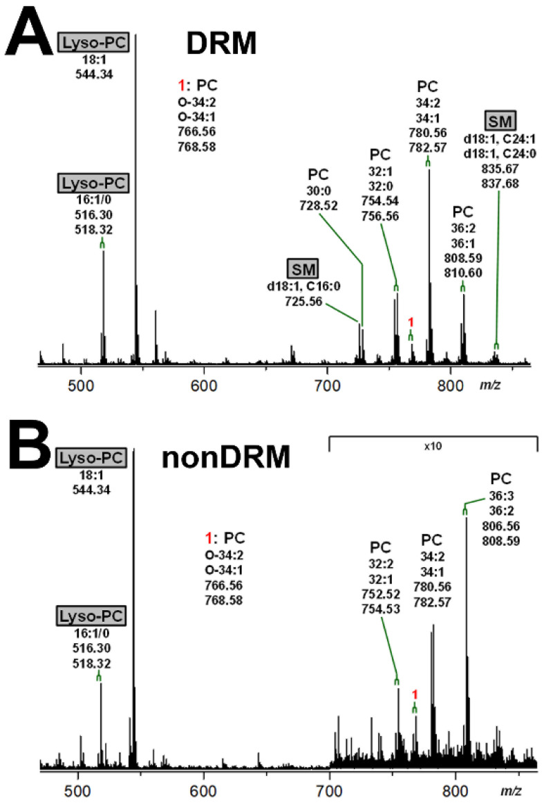
Figure 6.
MS1 spectra of phospholipids of DRM fraction F2 (A) and nonDRM fraction F7 (B) obtained from replicate 2 of pHRPTEpiCs. The spectra were recorded in the positive ion mode yielding monosodiated [M+Na]+ species, which could be assigned to the phospholipids indicated. SM species (gray boxes) highlight this characteristic marker of the liquid-ordered membrane phase, whereas lyso-PC species (gray boxes) appear in both the liquid-ordered (F2) and the liquid-disordered membrane phase (F7). PC, phosphatidylcholine; SM, sphingomyelin.

