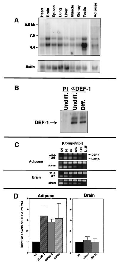
FIG. 6.
DEF-1 expression in various tissues and adipose tissue from mouse models of obesity. (A) The full-length DEF-1 cDNA was used to probe a mouse multiple-tissue Northern blot (Clontech), and 5 μg of poly(A)+ mRNA was isolated from mouse adipose tissue. The lower band appears to be smaller than the DEF-1 composite cDNA isolated and, therefore, is believed to be a DEF-1-related mRNA (data not shown). A β-actin probe was subsequently used to normalize mRNA loading. (B) Lysates from 3T3 F442A cells before (Undiff.) and after (Diff.) differentiation with 1 μM pioglitazone were immunoprecipitated with preimmune (PI) or anti-DEF-1 antiserum (αDEF-1). The immunoprecipitates were immunoblotted with the anti-DEF-1 antibody. (C) An example of the data obtained from a competitive PCR analysis used to evaluate the levels of DEF-1 mRNA in adipose and brain tissues isolated from wild-type, ob/ob, and db/db mice. The amount of competitor added that resulted in equivalent DEF-1 and competitor signal intensities was determined. (D) The average fold increase of DEF-1 mRNA levels in adipose tissue isolated from two ob/ob mice and one db/db mouse relative to that of a wild-type standard was determined as described in panel C. A similar analysis was performed with RNA from brain tissue. All values represent the average from three separate trials, and the error bars refer to average deviations.

