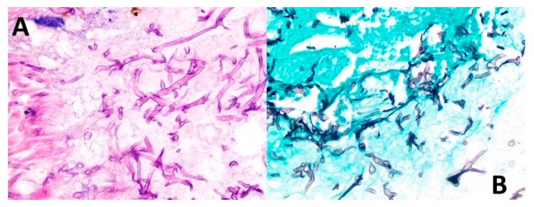
Figure 6.
Biopsy findings. (A) Routine staining with hematoxylin and eosin shows broad ribbon-shaped hyphae without marked septa, 5–15 microns in diameter and irregular angled branching ranging from 45 to 90 degrees. (B) Grocott’s methenamine stain permeates the walls of fungal cells due to its high density of polysaccharides, retaining more aldehyde groups by oxidation of chromic acid and highlighting the hyphal structures.

