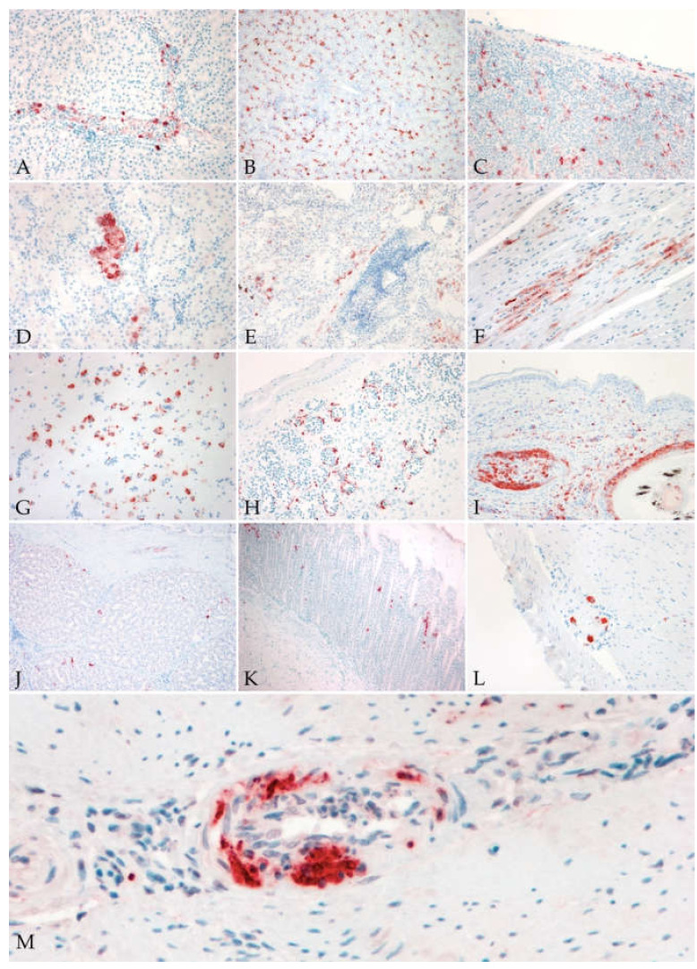
Figure 2.
Virus antigen in different tissues of blackbirds naturally infected with USUV. (A) Liver: positive intravascular mononucleated cells (20×); (B) liver: diffuse presence of virus antigen in Kupffer cells and hepatocytes (10×); (C) spleen: diffuse distribution of virus antigen in mononucleated cells and in smooth muscle and fibroblast of the splenic capsule (10×); (D) kidney: focal positivity of tubular epithelium (40×); (E) lung: multifocal positive pneumocytes, mononucleated inflammatory cells and smooth muscle cells of a pulmonary vessel (10×); (F) heart: focal positivity of cardiomyocytes; (G) cerebrum: diffuse positivity of neurons and glial cells (40×); (H,I) skin: diffuse positivity of endothelial cells (H; 20×) and positive feather shafts and bulbs (I; 20×); (J) proventriculus: scattered positive mucosal epithelial cells (10×); (K) gizzard: multifocal areas with positive mucosal epithelial cells (10×); (L) intestine: positive ganglionic neurons of myenteric plexus (20×); (M) medium size artery: positive signal in endothelial cells and in the infiltrating inflammatory cells (vasculitis) (40×).

