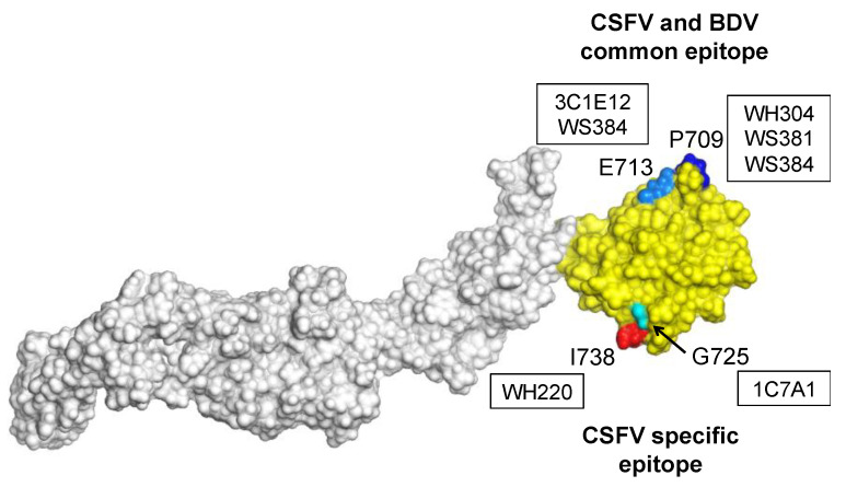
Figure 4.
The 3D structure modeling of E2 of CSFV strain TD/96. The domain B/C is colored yellow. The residues P709, E713, G725 and I738 recognized by mAbs are highlighted with dark blue, light blue, light green and red, respectively. The mAbs recognizing the highlighted amino acid residues are indicated in boxes.

