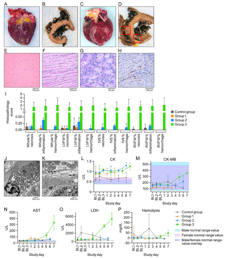
Figure 4.
Cowpox virus (CPXV) directly infects cardiac tissue. (A,B) Gross pathology image of a fresh, normal heart and formalin-fixed and transversely sectioned normal heart tissue (control group macaques). (C,D) Petechiae and areas of hemorrhage and necrosis (Group 3 macaques). (E,F) Normal control group and Group 3 ventricular myocardium section with florid inflammation. (G) Higher magnification of F showing intracytoplasmic inclusion bodies and inflammatory infiltrate. (H) CPXV antigen (brown) in cardiac fibroblasts. (I) Histopathology scoring. (J,K) Normal control group and Group 3 myocardium. Asterisk: mitochondrial crystolysis; arrows: CPXV particles in cardiac fibroblast. (L–P) Creatine kinase (CK), CK myocardial band (CK MB), aspartate aminotransferase (AST), lactate dehydrogenase (LDH), and hemolysis over time. BL = baseline sampling, time points prior to intravenous (IV) CPXV exposure. Day 0 is not included on the graphs because no measurements were made.

