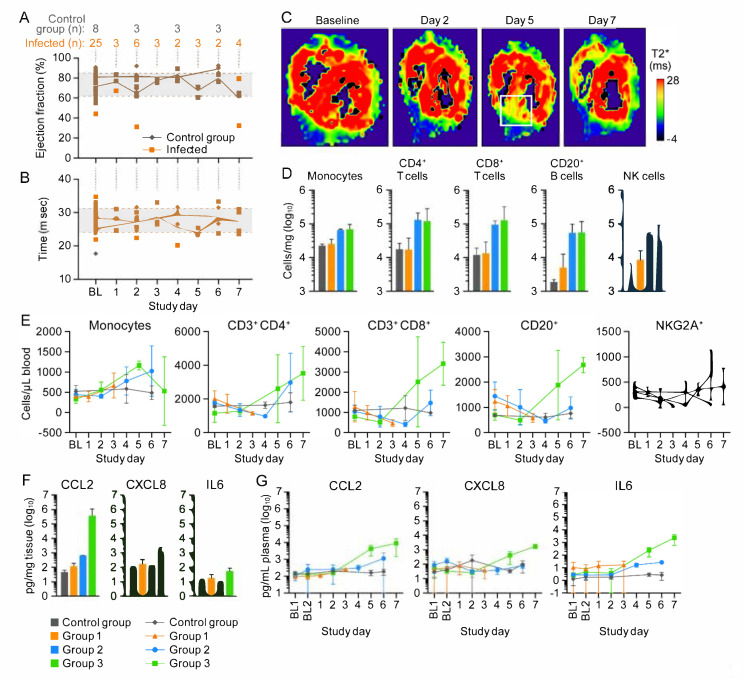
Figure 5.
Myocardial hemorrhage and lymphohistiocytic inflammation in cowpox-virus-exposed macaques does not severely impact heart function. (Baseline values were averaged to establish a mean and one standard deviation left ventricular ejection fraction [LVEF] normal range.) (A) Changes in LVEF. (B) Myocardial hemorrhage as measured by T2*. (C) T2* maps of the most severely affected macaque. White square: area with increased T2*, indicating hemorrhage. (D) Peripheral blood mononuclear cells (PBMCs) infiltrated the heart tissue and peak at Day 6 post-exposure. (E) Circulating PBMC numbers increased by Day 4. Monocyte numbers declined in circulation by Day 7 but remained constant within the myocardium. (F,G) Heart tissue and plasma cytokine concentrations increased as disease progressed. Day 0 is not included on the graphs because no measurements were made.

