Figure 6.
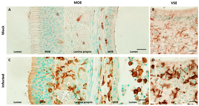
Inflammatory cells infiltrated olfactory structures. Scale bars = 20 µm. (A) DAB staining with the antibody against Iba1 in the MOE of the mock group. Iba1-positive cells were mostly present in the lamina propria and some were adjacent to the basal lamina; (B) DAB staining with the antibody to Iba1 in the VSE of the mock group. Iba1-positive cells showed their processes, which meant that they were in a resting state; (C) DAB staining with the antibody against Iba1 in the MOE of the infected group. There were increased Iba1-positive cell levels. These cells were located in the lamina propria, a relatively superficial portion of the epithelia, and the lumen; (D) DAB staining with the antibody to Iba1 in the VSE of the infected group. Many Iba1-positive cells were amoeboid in shape and more densely stained. They were in an active state.

