Figure 4.
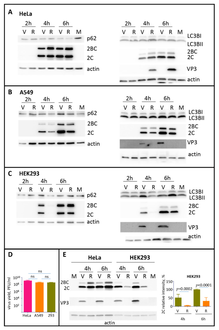
The development of autophagy upon polio infection is cell type specific. (A–C) HeLa, A549, or HEK293 cells, respectively, were infected with the MOI of 25 of either poliovirus (V) or encapsidated P2P3 replicon (R), and the cells were lysed at the indicated times post-infection. Lysates from mock-infected cells were prepared at 6 h. The same lysates were resolved either on 12% (left panels) or on 4–15% gradient gels (right panels) and analyzed in Western blots with the indicated antibodies. Staining with anti-2C antibody shows polio replication, staining with anti-VP3 antibody confirms the absence of capsid protein expression in cells infected with the replicon construct, actin is shown as a loading control. (D) HeLa, A549 and HEK 293 cells were infected with 25PFU of poliovirus and the virus yield at 6 h p.i. was determined by plaque assays. The difference in the titers between any of the samples was non-significant (ns). (E) HEK293 and HeLa cells were infected in parallel with the same preparations of either poliovirus (V) or encapsidated P2P3 replicon (R), collected at the indicated times post-infection and analyzed in a Western blot with anti-polio 2C and VP3 (capsid protein) antibodies. Actin is shown as a loading control. Note a significantly compromised replication of replicon compared to poliovirus in HEK293 but not HeLa cells. Quantitation shows 2C signals normalized to that in the virus-infected sample at 6 h p.i. from three independent experiments of infection of HEK293 cells with encapsidated replicon and poliovirus.

