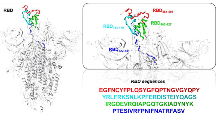
Figure 5.
Illustration of the RBD S-protein main chain as a gray carbon cartoon. Right-hand images show zoomed-in context of the four sequences inducing RBD-specific antibodies, as proposed in the present study, which were coloured as red (RBD484–508), cyan (RBD453–476), green (RBD402–427), or blue (RBD322–341) main chain ribbons.

