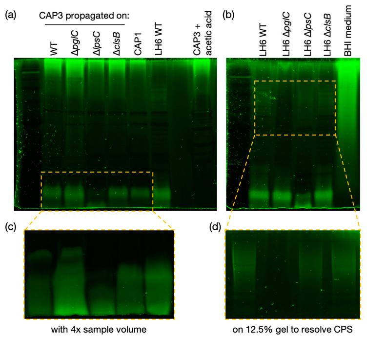
Figure 7.
Pro-Q™ Emerald 300 staining of CAP3 glycans. (a) Proteinase K digests of CAP3 propagated on LH6 WT, ∆pglC, ∆lpsC and ∆clsB, and CAP1 phages were separated on a 15% SDS-PAGE gel and stained with Pro-Q™ Emerald 300 glycan staining kit (compare with Figure S5). CAP3 was treated with 1% acetic acid to test whether the low molecular weight glycan is a lipid-linked glycoconjugate (and Figure S6). (b) Proteinase K digests of LH6 WT, ∆pglC, ∆lpsC and ∆clsB were separated on a 15% SDS-PAGE gel and stained to show differences in glycosylation. BHI medium was also tested to determine the origin of the high molecular weight glycan in the phage samples. (c) CAP3 propagated on LH6 WT, ∆pglC, ∆lpsC and ∆clsB, and CAP1 phages were loaded with 4x volume (from Figure S6). (d) Proteinase K digests of LH6 WT, ∆pglC, ∆lpsC and ∆clsB were separated on a 12.5% SDS-PAGE gel to better resolve high molecular weight capsular polysaccharides (from Figure S7).

