Figure 2.
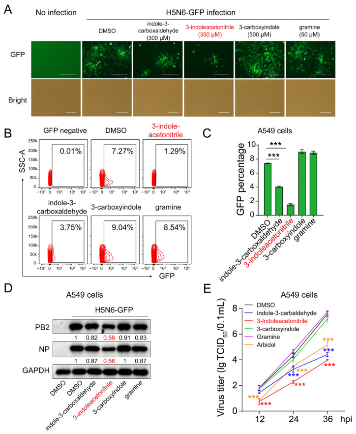
The effect of indole derivatives on H5N6-GFP virus replication. The A549 cells were infected with the H5N6-GFP virus at an MOI of 0.005, followed by treatment with 3-indoleacetonitrile, indole-3-carboxaldehyde, 3-carboxyindole, and gramine at indicated concentrations for 24 h. After that, the GFP intensity was acquired using fluorescence microscopy (A). The percentage of GFP-positive cells was calculated through flow cytometry (B, a presentative image) (C, data collected from three independent biological experiments). The viral PB2 and NP proteins were analyzed by Western blotting (D). The growth curves of H5N6-GFP virus in supernatants were determined based on three time points of infection as indicated (E), 3-indoleacetonitrile (350 μM); indole-3-carboxaldehyde (300 μM); 3-carboxyindole (500 μM); gramine (50 μM); arbidol (8 μM). *** p < 0.001; calculated from three independent experiments by two-tailed Student’s t-test (C) or two-way ANOVA (E).

