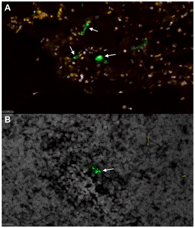
Figure 3.
Fluorescent immunohistochemistry for SARS-CoV-2 nucleoprotein identifies mononuclear cells in tracheobronchial lymph nodes of intratracheally infected cats. Low numbers of SARS-CoV-2 positive cells (green, white arrows) are detected in (A) positive control tissue (lung) from an African Green Monkey infected with SARS-CoV-2 [50], and within (B) mononuclear cells in the TBLN of SARS-CoV-2-infected cats (green, white arrow). White, DAPI/nuclei; green, CoV-2. Magnification 40×, scale bar = 20 µm.

