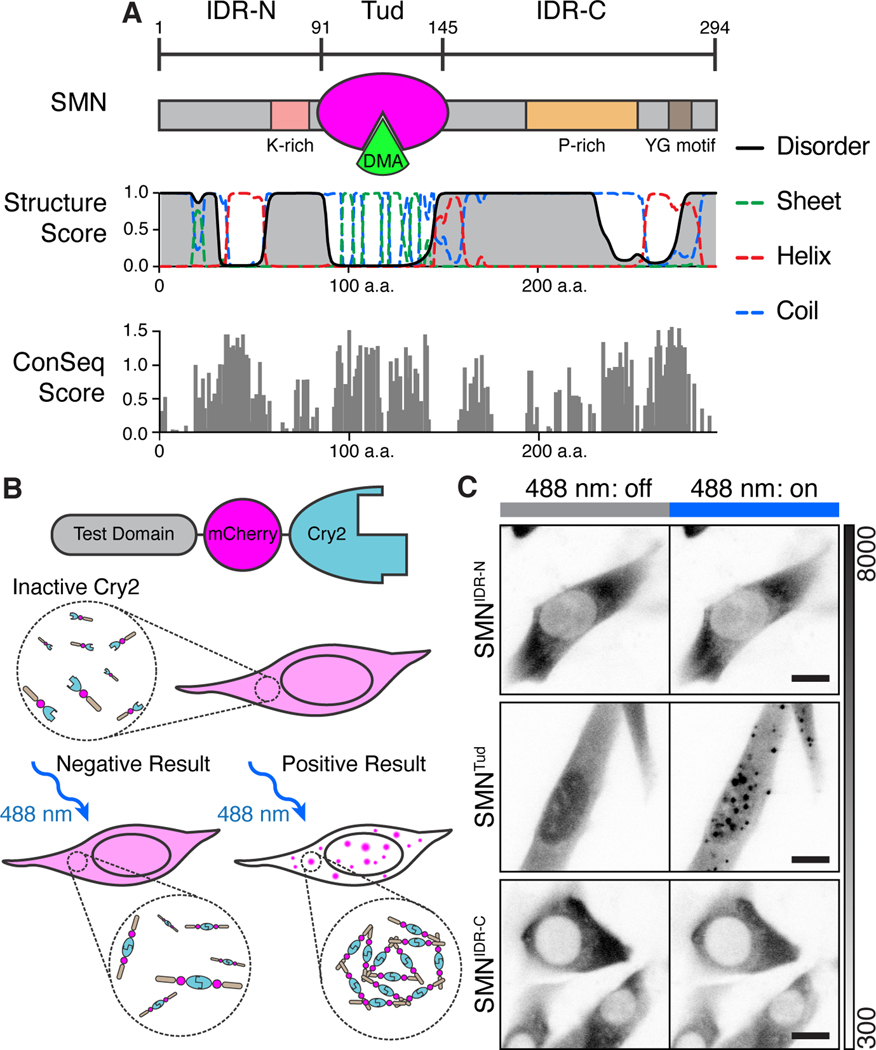
Figure 1. The multimerized SMN tudor domain forms condensates in vivo.
A) Schematic representation of SMN domain architecture and accompanying secondary structure prediction. The tudor (Tud, magenta) domain binds DMA (green). Structure score is a unitless value for secondary structural properties predicted by the RaptorX algorithm. ConSeq scores are displayed for SMN residues aligned using Clustal Omega and computed using ConSeq (Berezin et al., 2004). The absolute value of conservation scores for the 50% most conserved residues are displayed. B) Diagram of the “optodroplet” condensation assay. Cry2 dimerizes upon blue light activation (488 nm); without added molecular interaction contributed by the test domain, condensation will not occur (Shin et al., 2017). If the test domain provides interactions to increase valency, condensation is observed as mCherry fluorescent foci. C) Micrographs of live cells undergoing blue light activation of Cry2 (180 s, blue bar). Grayscale bar given in analog-digital units. Scale bar = 10 μm.

