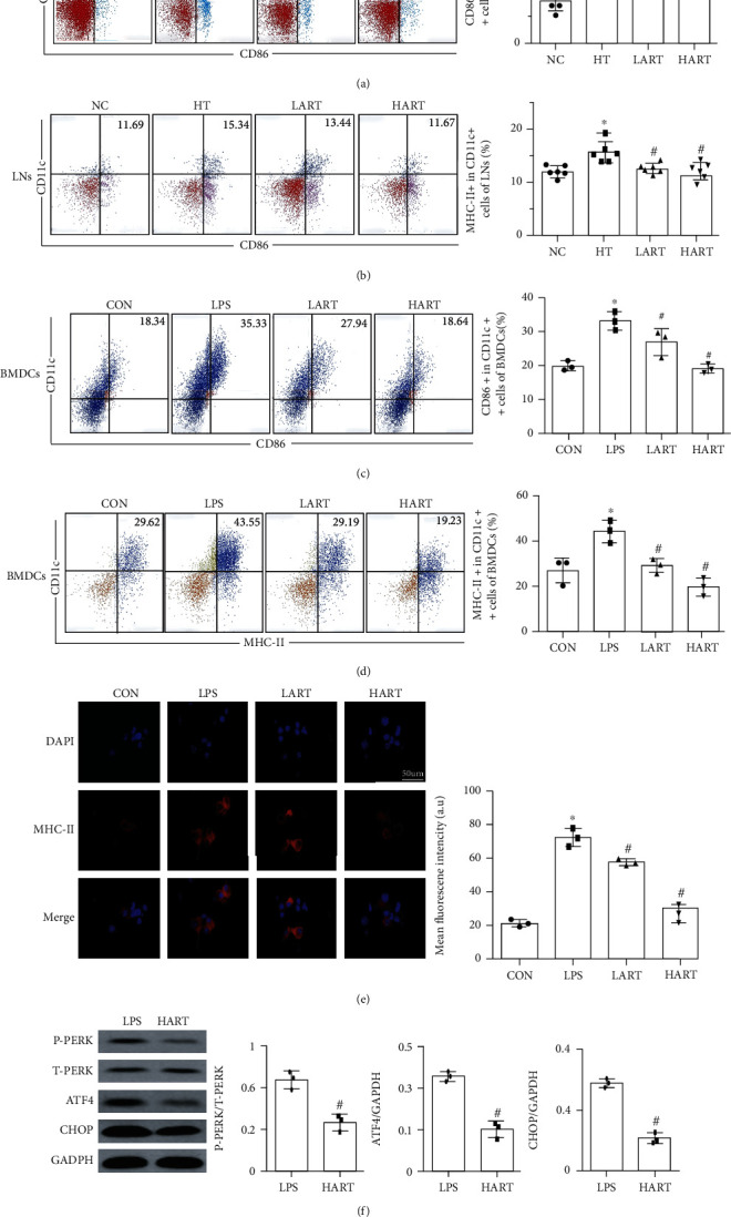
Figure 5.

(a) Flow cytometric sorting of CD11c+ and CD86+ cells in lymph nodes; (b) flow cytometric sorting of CD11c+ and MHC-II+ cells in lymph nodes (∗P < 0.05 compared with NC; #P < 0.05 compared with HT); (c) flow cytometric sorting of CD11c+ and CD86+ cells in BMDCs; (d) flow cytometric sorting of CD11c+ and MHC-II+ cells in BMDCs (∗P < 0.05 compared with CON; #P < 0.05 compared with LPS); (e) immunofluorescence staining and mean fluorescence intensity of MHC-II in BMDCs (blue: DAPI, red: MHC-II); (f) western blotting of PERK/ATF4/CHOP signaling pathway (#P < 0.05 compared with LPS).
