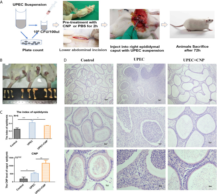
Figure 1.
Morphological traits of acute caput epididymitis in rats. (A) Steps to establish the model of epididymitis. (B) Visual changes of caput epididymitis and rat epididymal index. (C) The CNP content of caput epididymal fluid was determined by ELISA. (D) H&E staining showed the characteristics of rat caput epididymitis under a microscope. Bar = 50 um. The data shown are representative of multiple experiments (mean ± SD, n = 5, *P < 0.05, **P < 0.01, ***P < 0.001).

