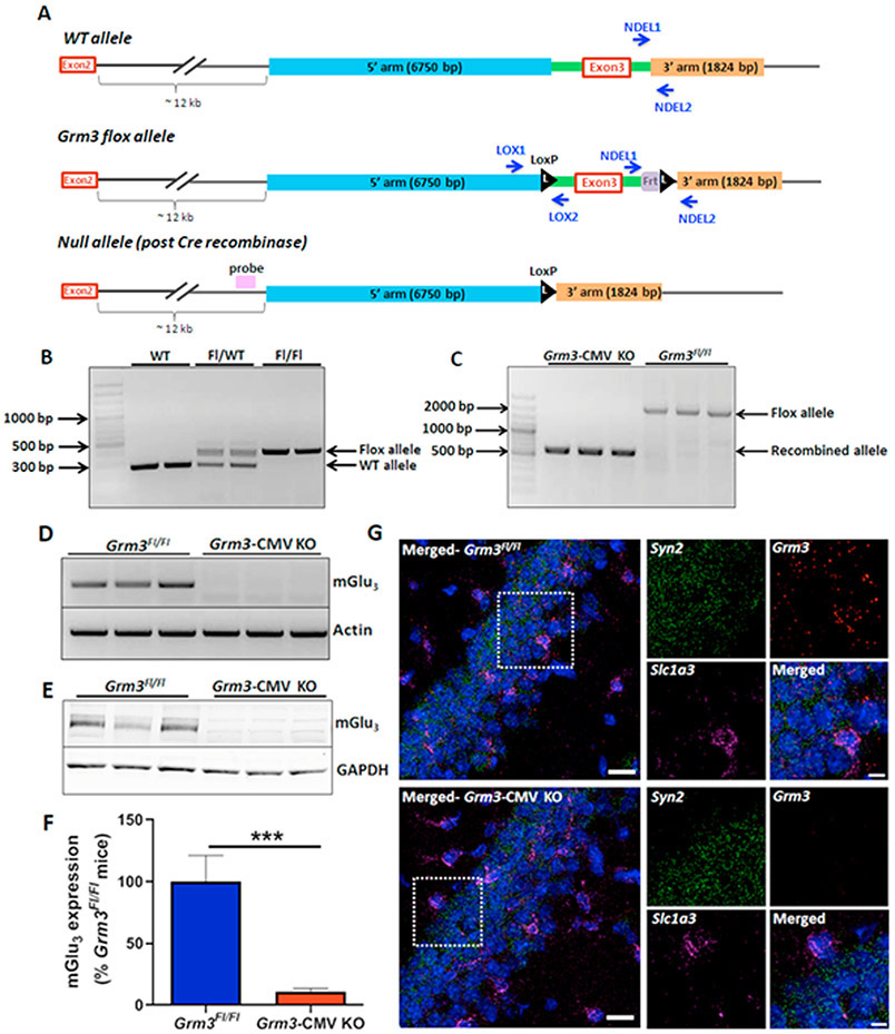
Figure 6. Generation and characterization of conditional Grm3Fl/Fl mice.
(A) Schematic of the procedure for generating the floxed Grm3 clones. (Top) Wild-type Grm3 locus surrounding exon 3. (Middle) Floxed Grm3 locus where LoxP sites are inserted flanking exon 3 of Grm3. (Bottom) Cre-mediated recombination leading to loss of Grm3 exon 3 and Frt site. (B) PCR from the DNA of mice homozygous for the WT allele (277 bp) and mice heterozygous and homozygous for the floxed allele (413 bp). (C) CMV-Cre mediated recombination in Grm3Fl/Fl mice excises Grm3 exon 3, leading to loss of WT allele (~1900 bp) and generation of a smaller recombination allele (671 bp). (D) RT-PCR from the hippocampi of WT and Grm3-CMV KO mice. (E) Western blot depicting loss of mGlu3 protein from the hippocampus of Grm3-CMV KO mice (red bar) relative to Grm3Fl/Fl controls (blue bar). (F) Bar graph depicting quantification of Western blots. Data are presented as mean ± SEM, N=3-6 mice. (***p<0.001 compared to Grm3Fl/Fl mice, t(7)= 7.018, Student’s t-test). (G) Confocal 40X RNAscope in situ hybridization images showing loss of Grm3 mRNA (red) from both neurons (Syn2; Synapsin, green) and astrocytes (Slc1a3; GLAST, magenta) in Grm3-CMV KO mice. Scale bar = 20 μm for the merged left panel image, and 10 μm for the 3X images.

