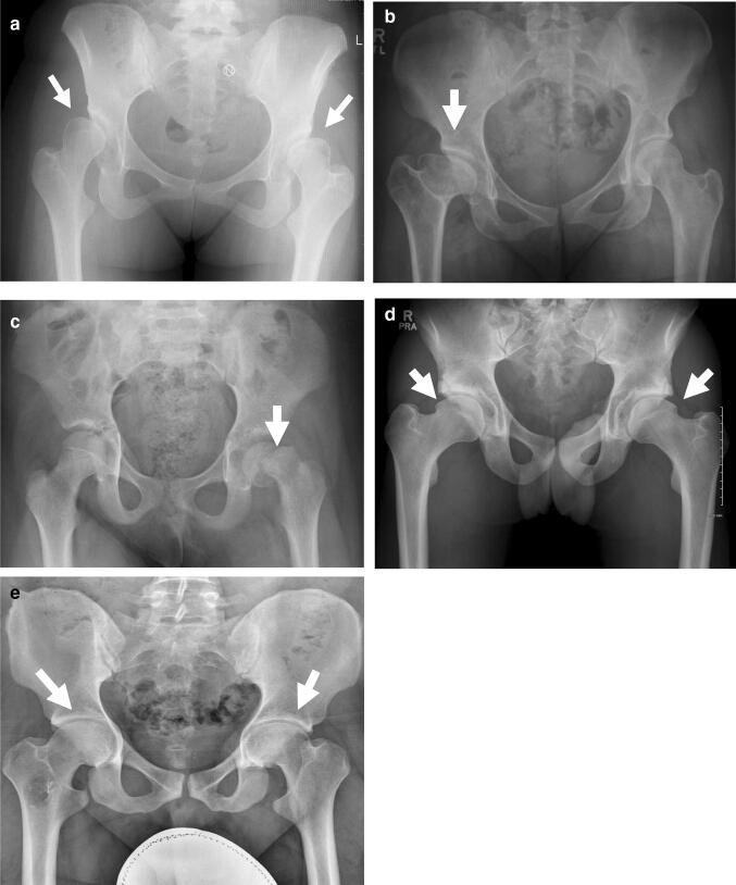
Fig. 3.
Disorders of the growing hip joint. a Bilateral hip dysplasia in a skeletally mature individual. The acetabuli are typically shallow and steep, and there is extrusion of the femoral heads with evidence of decreased lateral coverage (arrows); b Perthes’ disease of the right hip in a skeletally mature individual. The right femoral head is broad and flattened and the corresponding acetabululm is similarly shaped (arrow). c Slipped capital femoral epiphysis of the left hip in an adolescent male. The epiphysis has slipped posteromedially from the physis (arrow); d Bilateral cam lesions in a skeletally maturing male, arrows show lateral head-neck junction prominence; e Bilateral pincer lesions in a skeletally mature male, arrows show over-coverage of femoral head

