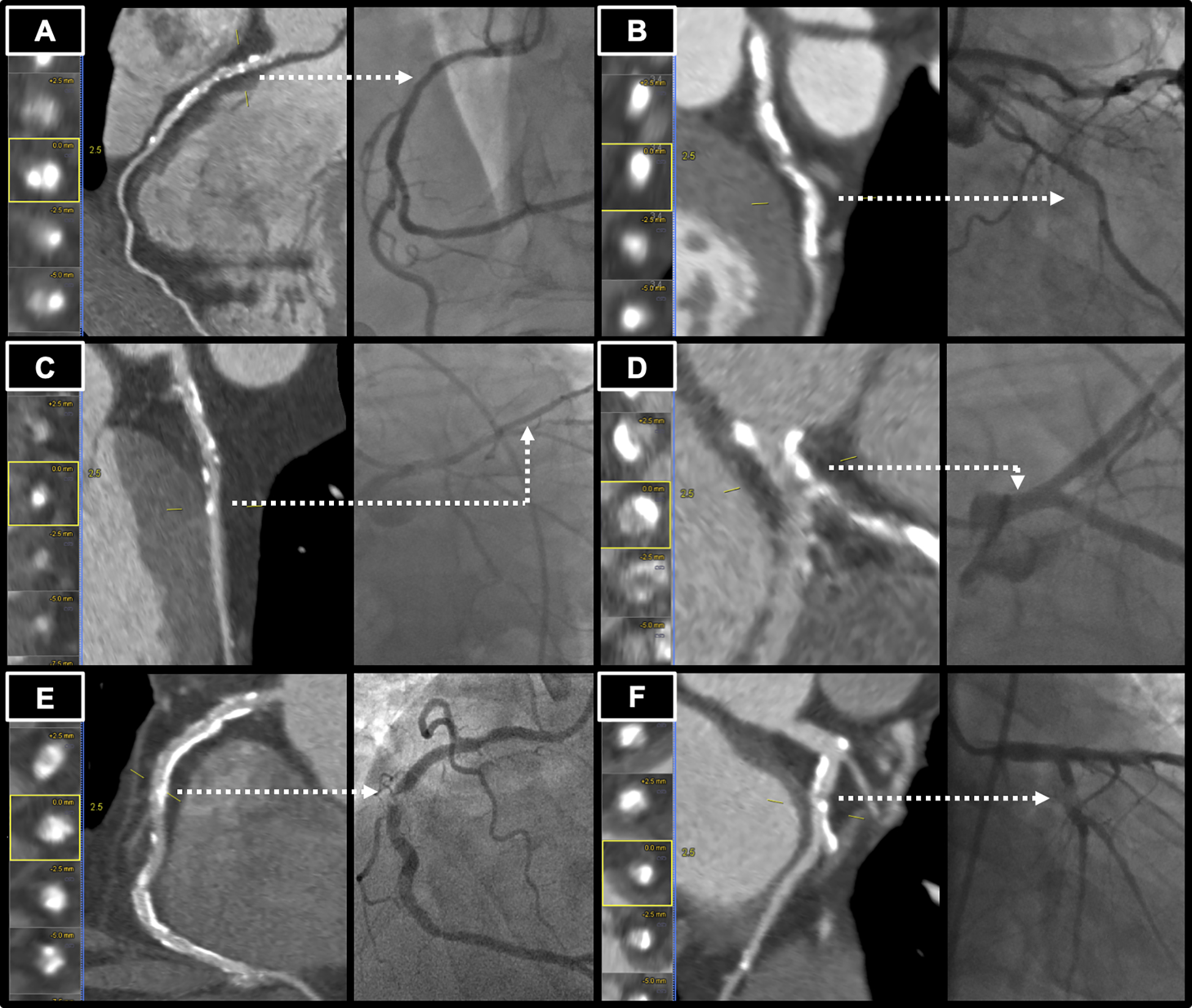
Figure 2.

Examples of calcified lesions resulting in incorrect estimate by coronary CT angiography (CCTA). Dashed arrow indicates same lesion in CCTA and cardiac catheterization. (A) Left Panel: Calcified stenosis in the proximal right coronary artery (RCA) graded as 70–99% stenosis by CCTA. Right Panel: Cardiac catheterization view with 46% stenosis by quantitative coronary angiography (QCA). (B) Left Panel: Calcified stenosis in the left circumflex artery (LCx) graded as 70–99% stenosis by CCTA. Right Panel: Cardiac catherization view with 11% stenosis by QCA. (C) Left Panel: Calcified stenosis in the left anterior descending artery graded as 70–99% stenosis by CCTA. Right Panel: Cardiac catherization view with 32% stenosis by QCA. (D) Left Panel: Calcified stenosis in the left main coronary artery graded as 50–69% stenosis by CCTA. Right Panel: Cardiac catherization view with 6% stenosis by QCA. (E) Left Panel: Calcified stenosis in the mid RCA, graded as 25–49% stenosis by CCTA. Right Panel: Cardiac catheterization view with 97% stenosis by QCA. (F) Left Panel: Calcified stenosis in the LCx graded as 25–49% stenosis by CCTA. Right Panel: Cardiac catherization view with 93% stenosis by QCA.
