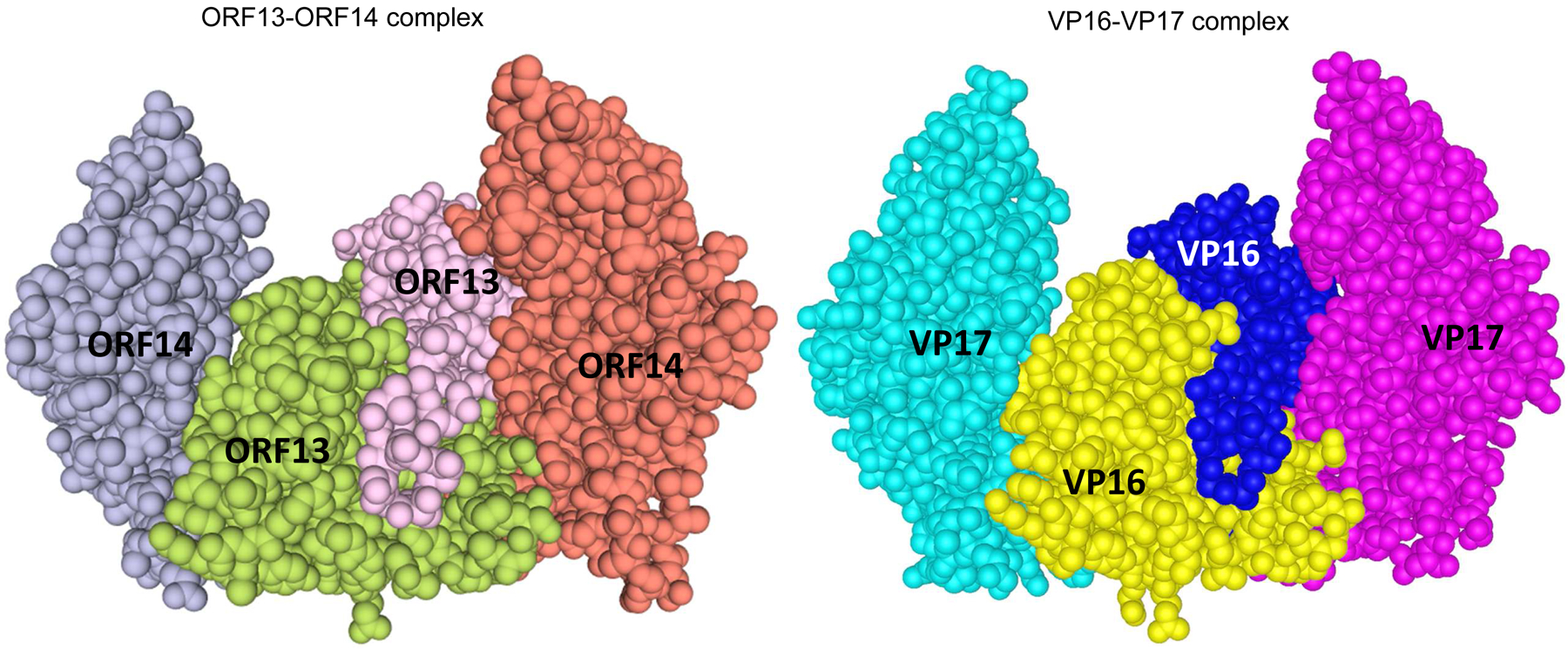
Figure 4:

Three dimensional (3D) structures of coat proteins of bacteriophages ΦIN93 and P23–77. The 3D structure of ORF13-ORF14 complex (left) from bacteriophage ΦIN93 was modeled, based on sequences and the 3D structure of VP16–VP17 complex (right; Cn3D view) from bacteriophage P23–77 as template, using Swiss-Model software. Homodimers of ORF13 (middle) and VP16 (middle) interact with each of the two copies of ORF14 and VP17, respectively. Both structures were modeled using amino acids 21–165 from ORF13 or VP16 and amino acids 46–271 from ORF14 or VP17 (highlighted in gray-background in Figure 1).
