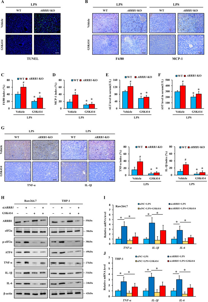
Fig. 6. Blockage of ER stress abrogated ARRB1-modulated activation of liver macrophages in vivo and in vitro.
Mice were intraperitoneally administered LPS injection (5 mg/kg) 8 h after gavage with GSK414 (150 mg/kg) or vehicle solution. n = 6 in each group. A TUNEL staining of livers in LPS-induced mice with or without GSK414 challenge (scale bar: 100 μm). B–D F4/80 and MCP-1 staining of livers in LPS-induced mice with or without GSK414 challenge (scale bar: 50 μm), and the positive F4/80 and MCP-1 indexes were scored. E, F Serum ALT and AST levels in LPS-induced mice with or without GSK414 challenge. G TNF-α and IL-1β staining of livers in LPS-induced mice with or without GSK414 challenge (scale bar: 50 μm), and the positive TNF-α and IL-1β indexes were scored. C–G *P < 0.05 compared with the respective vehicle treatment; #P < 0.05 compared with LPS-induced WT mice by Student’s t test. H–J Raw264.7 and THP-1 cells were challenged with LPS (1 μg/ml) for 6 h with or without GSK414 (1 μM) treatment after knockdown of ARRB1 by siRNA. H Western blotting analysis of the indicated protein expression. I, J TNF-α, IL-1β, and IL-6 mRNA levels in Raw264.7 and THP-1 cells in the indicated treatments were analyzed by real-time PCR. *P < 0.05 by Student’s t test. Data are presented as the mean ± SD of three separate experiments.

