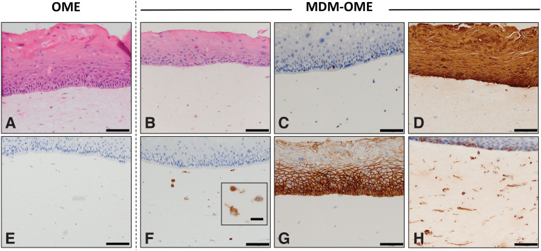
FIG. 2.
Histological characterization of MDM-OME. Models were cultured for 10 days at air-to-liquid interface and analyzed by histology with hematoxylin and eosin staining (A, B) and immunohistochemistry for immunopositive expression of ki67 (C) cytokeratin (AE1/3) (D), CD68 (E, F), E-cadherin (G), and vimentin (H). Images presented as representative from at least three independent experiments. Scale bar = 100 μm and 20 μm for the inlay box in (F). OME, oral mucosa equivalent. Color images are available online.

