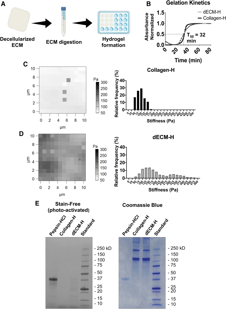
FIG. 2.
dECM-H formation and characterization. (A) General schematic of the derivation of dECM. (B) Gelation kinetics of dECM-H and Collagen-H, where t50 is the time it takes for dECM-H to reach 50% of maximum absorbance. (C) Force maps generated from AFM and the relative frequency of stiffness seen in Collagen-H. (D) Force maps generated from AFM and the relative frequency of stiffnesses seen in dECM-H. (E) SDS-PAGE results on a stain-free gel, which was then stained further with Coomassie Blue. AFM, atomic force microscopy; Collagen-H, collagen type I hydrogel; dECM-H, dermis extracellular matrix hydrogel; SDS-PAGE, sodium dodecyl sulfate–polyacrylamide gel electrophoresis. Color images are available online.

