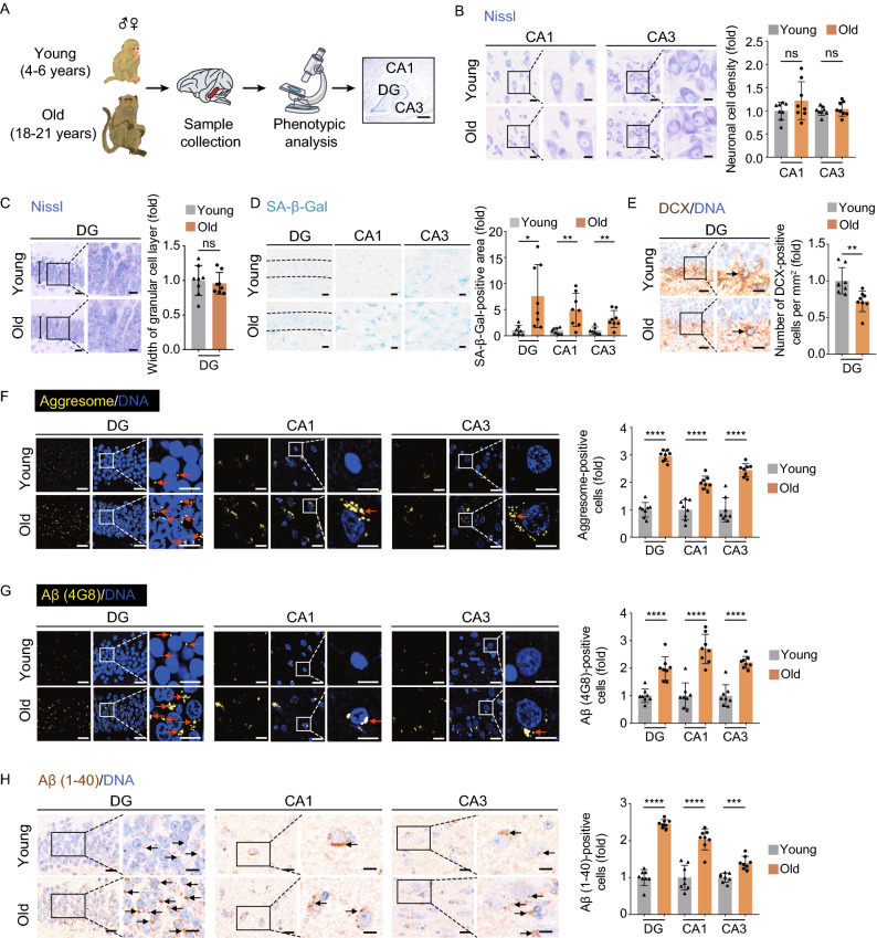
Figure 1.
Aging-related phenotypes of the cynomolgus monkey hippocampus. (A) Flow chart of the phenotypic analysis on the hippocampal tissues collected form young and old monkeys. Representative image of Nissl staining in the hippocampus is shown (right panel, dentate gyrus (DG), CA1 region (CA1) and CA3 region (CA3), scale bar, 700 μm). (B) Nissl staining in CA1 and CA3 regions of the hippocampus from young and old monkeys. Representative images are shown on the left; neuronal cell densities in corresponding regions are quantified as fold changes (old vs. young), shown as means ± SEM on the right. Scale bars, 20 μm and 10 μm (zoomed-in image). Young, n = 8; old, n = 8 monkeys. ns, not significant. (C) Nissl staining in the dentate gyrus (DG) from young and old monkeys. Representative images are shown on the left; the widths of the granular cell layers in the DG are quantified as fold changes (old vs. young), shown as means ± SEM on the right. Scale bars, 20 μm and 10 μm (zoomed-in image). Young, n = 8; old, n = 8 monkeys. ns, not significant. (D) SA-β-Gal staining in the indicated regions of the hippocampus from young and old monkeys. Representative images are shown on the left; SA-β-Gal positive areas in the DG, CA1 and CA3 regions are quantified as fold changes (young vs. old), shown as means ± SEM on the right. Scale bar, 20 μm. Young, n = 7; old, n = 8 monkeys. *P < 0.05; **P < 0.01. (E) Immunohistochemical staining of DCX in the DG region of the hippocampus from young and old monkeys. Representative images are shown on the left; DCX-positive cells are quantified as fold changes of their numbers in the old DG vs. in young counterparts, shown as means ± SEM on the right. Black arrows indicate the DCX-positive cells. Scale bars, 20 μm and 10 μm (zoomed-in image). Young, n = 8; old, n = 8 monkeys. **P < 0.01. (F) Aggresome staining in the indicated regions of the hippocampus from young and old monkeys. Representative images are shown on the left; aggresome-positive cells are quantified as fold changes of their numbers in the old DG, CA1 and CA3 regions vs. in young counterparts, shown as means ± SEM on the right. Red arrows indicate the aggresome-positive cells. Scale bars, 20 μm and 10 μm (zoomed-in image). Young, n = 8; old, n = 8 monkeys. ****P < 0.0001. (G) Immunofluorescence staining of Aβ (4G8) accumulation in the indicated regions of the hippocampus from young and old monkeys. Representative images are shown on the left; Aβ (4G8)-positive cells are quantified as fold changes of their numbers in the old DG, CA1 and CA3 regions vs. young counterparts, shown as means ± SEM on the right. Red arrows indicate the Aβ (4G8)-positive cells. Scale bars, 20 μm and 10 μm (zoomed-in image). Young, n = 8; old, n = 8 monkeys. ****P < 0.0001. (H) Immunofluorescence staining of Aβ (1-40) accumulation in the indicated regions of the hippocampus from young and old monkeys. Representative images are shown on the left; quantitative data for the relative Aβ (1-40)-positive cells in the DG, CA1 and CA3 regions are shown as means ± SEM on the right. The relative fold of number of Aβ (1-40)-positive cells was obtained by normalizing the number of Aβ (1-40)-positive cells of the old monkey with the young monkey. Black arrows indicate the Aβ (1-40)-positive cell. Scale bars, 20 μm and 10 μm (zoomed-in image). Young, n = 8; old, n = 8 monkeys. ***P < 0.001, ****P < 0.0001

