Fig. 2.
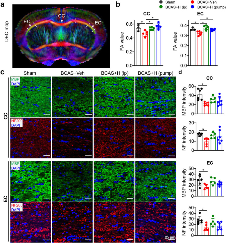
Blocking NHE1 with inhibitor HOE642 in BCAS mice ameliorated white matter damage. a Representative DEC maps showing corpus callosum (CC) and external capsule (EC) for DTI analysis. At 30 days post-BCAS following neurological function analysis in Fig. 1d, mice were sacrifized for ex vivo brain MRI DTI and subseqent immunoflurorescence staining. b Quantitative analysis of mean FA values in CC and EC. Data are presented as mean ± SD. sham group: n = 3, BCAS+Veh and BCAS+HOE (i.p. or pump): n = 5–6. *p < 0.05. c Representative images of MBP or NF staining in CC and EC. However, due to lack of optimal anti-MBP and NF200 antibodies for double labeling, the images in Fig. 2C were obtained from anti-MBP or anti-NF200 staining, respectively. d Quantitative analysis of immunofluorescent intensity of MBP and NF in CC and EC, respectively. Data are presented as mean ± SD. n = 5–6/group. *p < 0.05

