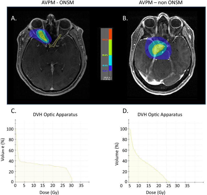
Fig. 2.
Radiation plan and Dose Volume Hisogram for AVPM patients with and without optic nerve sheath (ONSM) involvement. Both AVPM patients received hypofractionated radiosurgery (hSRT) in 5 sessions × 500 cGy, with maximum dose of 31.25 Gy. Optical apparatus (left and right optic nerve, optic chiasm) was marked in treatment plans of both patients and DVHs were calculated. A Radiation plan for Optic Nerve Sheath Meningioma (ONSM) patient. B Radiation plan for non-ONSM AVPM patient. Comparison of Optic apparatus DVH reveals that in the ONSM patient (C) a higher radiation dose was absorbed by the nerve sheath while in the perioptic non-ONSM AVPM patient (D) the same radiation dose to the tumor resulted in smaller dose to the nerve sheath. We believe the combination of pre-treatment nerve damage and the high dose to the nerve explain the increased visual damage we see in ONSM patients treated with hSRT and suggest this cohort should be treated with cFSRT

