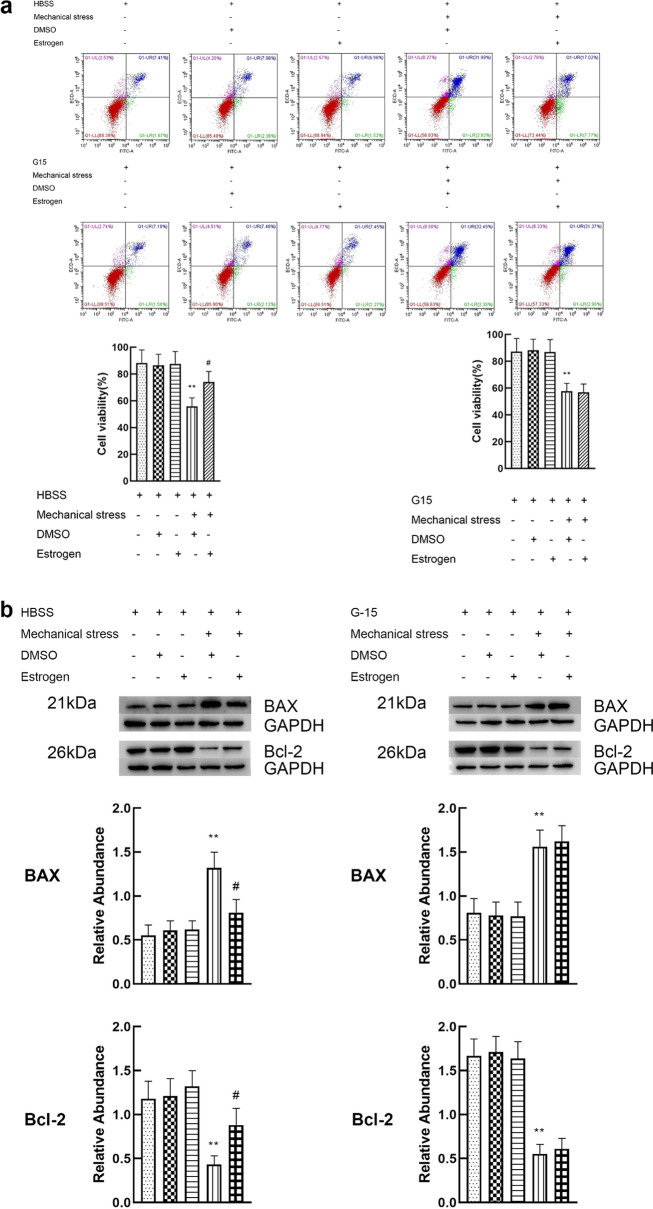
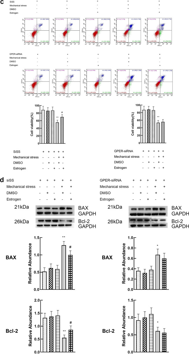
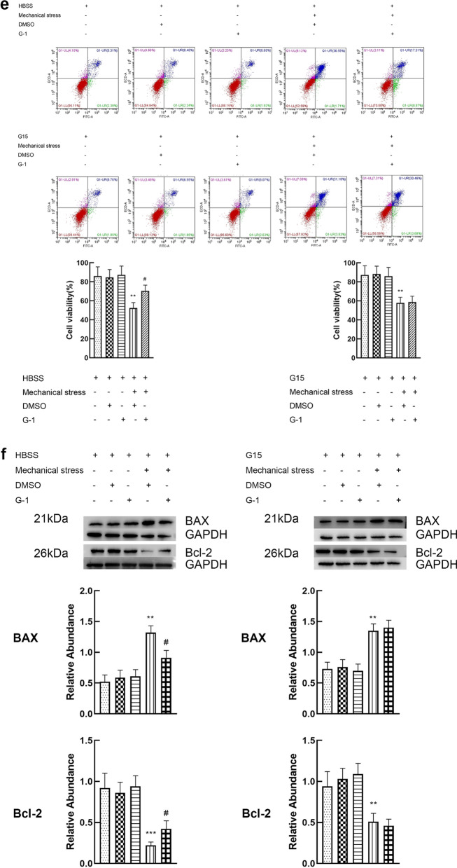
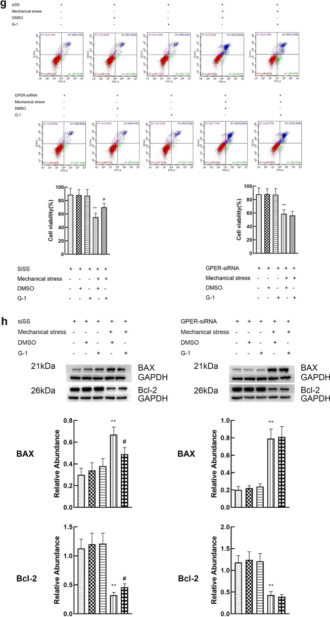
Fig. 3.
Activation of GPER reduces the apoptosis of chondrocytes induced by mechanical stress. a Cell viabilities for chondrocytes treated with mechanical stress, 8 μM estrogen, and 8 μM G15 (n = 3). b Protein expression of Bax and Bcl-2 for chondrocytes treated with mechanical stress, 8 μM estrogen, and 8 μM G15 (n = 3). c Cell viabilities for chondrocytes with GPER or scrambled siRNA after treatment with mechanical stress, and estrogen (n = 3). d Protein expression of Bax and Bcl-2 for chondrocytes with GPER or scrambled siRNA after treatment with mechanical stress, and estrogen (n = 3). e Cell viabilities for chondrocytes treated with mechanical stress, 10 μM G-1, and 8 μM G15. f Protein expression of Bax and Bcl-2 for chondrocytes treated with mechanical stress, 10 μM G-1, and 8 μM G15. g Cell viabilities for chondrocytes with GPER or scrambled siRNA after treatment with mechanical stress, and G-1 (n = 3). h Protein expression of Bax and Bcl-2 for chondrocytes with GPER or scrambled siRNA after treatment with mechanical stress, and G-1 (n = 3). All data are presented in mean ± SD (n = 3). *P < 0.05, compared with DMSO group. **P < 0.01, compared with DMSO group. #P < 0.05, compared with mechanical stress + DMSO group. ##P < 0.01, compared with mechanical stress + DMSO group


