Figure 1.

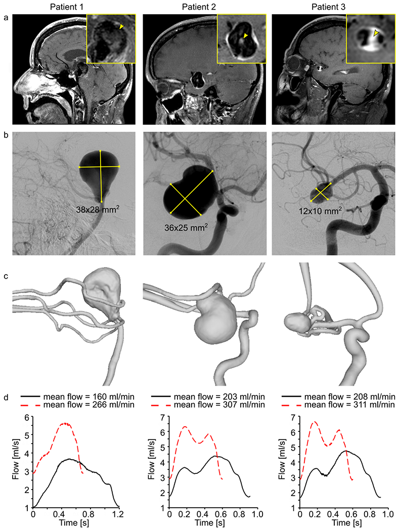
Black-blood MRI of patients 1-3 with intracranial aneurysm (yellow squares, inset) with wall-adjacent and luminal (yellow arrowhead) intra-aneurysmal signal enhancement after administering contrast agent (a), digital subtraction angiography (b), and surface rendering of segmented lumens (c) of intracranial arteries with aneurysms. Using these data, 3D-printed models were produced and supplied with the pulsatile flow at different velocities and cycles, measured with a sensor at the outlet (d).
