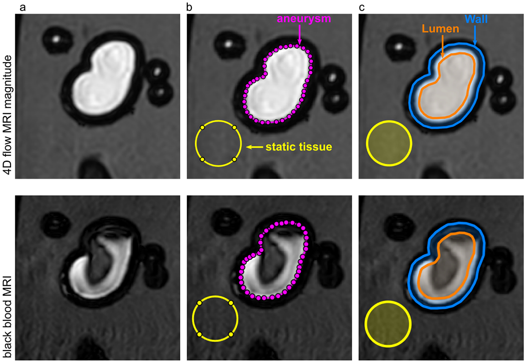
Figure 2.

4D flow magnitude MRI (top) and BB MRI (bottom) of model 2 with different regions of interest (ROI). ROIs for lumen (orange), wall (orange-blue), aneurysm (magenta), and static tissue (yellow) were manually created on the 4D flow MR magnitude images and translated to the black-blood MRI. The BB MRI signal obtained in the aneurysm ROI (magenta) and wall (orange-blue) was normalized by the signal of the static tissue (yellow) outside of the flow volume to calculate the signal enhancement on black-blood MRI.
