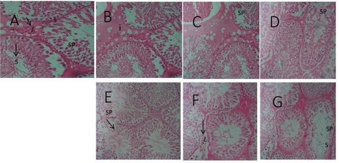
Figure 6 .

Testicular micrograph (H&E ×100): (A) Control; normal spermatogenic cells (SP), and no degeneration; (B) Alcohol; cells were normal with slight cell degeneration; (C) Tobacco cell were abnormal and degenerated; (D) Cannabis; normal cells and slightly abnormal cells; (E) Alcohol and Tobacco; morphological distorted cell and germinal cell degeneration; (F) Alcohol and Cannabis; cell morphological distortion and cell apoptosis; (G) Alcohol, Tobacco, and Cannabis; spermatogenic cells apoptosis and cells morphological distortion. Leydigs cell (L), seminiferous tubules (S).
