Fig. 1.
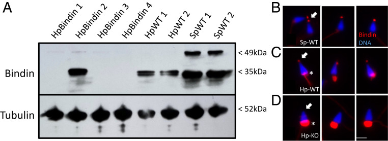
(A) Anti-bindin antibody reveals gene KOs in sperm. Sperm proteins were isolated, separated by sodium dodecyl sulfate polyacrylamide gel electrophoresis, blotted to nitrocellulose, and challenged by antibody incubation. Anti-tubulin antibodies were used to estimate relative loading levels of sperm protein. HpBindin represent sperm isolated from different individuals of bindin gRNA/Cas9 mRNA microinjections, whereas Hp-WT and Sp-WT are from WT sperm of Hp (Hp-WT) and S. purpuratus (Sp-WT). (B–D) Immunolabeling of sperm in situ with anti-bindin antibodies. (B) S. purpuratus sperm. Note the Bindin spot at the tip of the sperm (arrow). (C) Hp-WT sperm. Note Bindin at the tip of the sperm (arrow), and also background labeling of the midpiece (asterisk). Surprisingly, any intact IgG (containing an Fc region) binds to the midpiece in Hp, but the Fab used as secondary antibodies does not. (D) Hp bindin KO sperm. Note that there is no Bindin label at the tip of the sperm (arrow), even though background labeling of the midpiece is present (asterisk). (Scale bar, 4 μm.)

