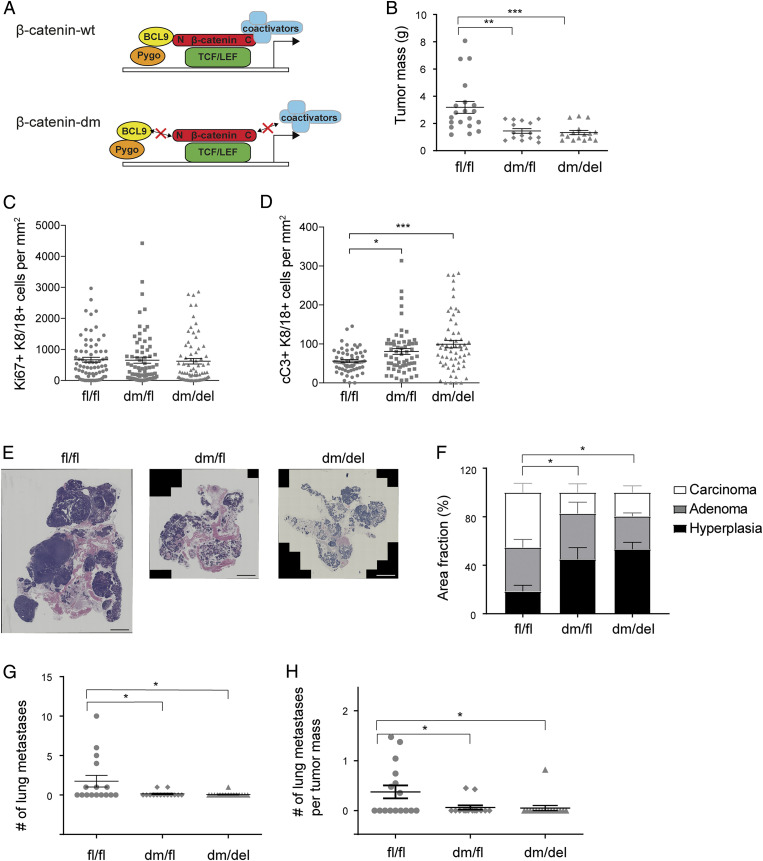
Fig. 2.
Loss of β-catenin cofactor binding impairs primary tumor growth, tumor progression, and metastasis formation. (A) Schematic representation of the mutant version of β-catenin used in this study. β-catenin can be divided into three distinct domains: an N-terminal region which binds Bcl9/9l, a central Armadillo (Arm) repeat region which binds TCF/LEF and E-cadherin, and a C-terminal region that binds several transcriptional coactivators. The dm version of β-catenin combines a D164A mutation in the first Arm repeat, which carries an amino acid change from aspartic acid to alanine, ablating the binding of Bcl9/9l to the N terminus, and a ΔC mutation, which carries a truncation of the C terminus of β-catenin, thereby preventing binding of C-terminal transcriptional coactivators. The red crosses on top of arrows highlight impaired interactions. It is important to note that the mutant form of β-catenin retains its cadherin-mediated cell adhesion function. (B) Primary tumor weights of mammary tumors of 12-wk-old MMTV-PyMT transgenic mice expressing in their mammary tumor cells wild-type (fl/fl) β-catenin, one wild-type and one mutant allele of β-catenin (dm/fl), or only a mutant allele of β-catenin (dm/del). Mouse numbers analyzed: fl/fl, n = 20; dm/fl, n = 15; dm/del, n = 16. The dots in the graphs represent the individual mice analyzed. Data are displayed as mean ± SEM. Statistical analysis was performed using ordinary one-way ANOVA multiple comparison test. **P < 0.01; ***P < 0.005. (C) Quantification of the immunofluorescence staining for Ki67 positive and K8/18 positive on tumor sections from β-cateninfl/fl;MMTV-PyMT (fl/fl), β-catenindm/fl;MMTV-PyMT (dm/fl), and β-catenindm/fl;MMTV-PyMT;MMTV-Cre (dm/del) mice at 12 wk of age. n = 5 mice, 12 imaging fields per mouse. Dots in the graphs represent the individual imaging fields analyzed. Data are displayed as mean ± SEM. (D) Quantification of the immunofluorescence staining for cC3 positive and K8/18 positive on tumor sections from β-cateninfl/fl;MMTV-PyMT (fl/fl), β-catenindm/fl;MMTV-PyMT (dm/fl), and β-catenindm/fl;MMTV-PyMT;MMTV-Cre (dm/del) mice at 12 wk of age. n = 5 mice, 12 imaging fields per mouse. The dots in the graphs represent the individual imaging fields analyzed. Data are displayed as mean ± SEM. Statistical analysis was performed using ordinary one-way ANOVA multiple comparison test. *P < 0.05; ***P < 0.005. (E) Representative images of histological tumor sections from β-cateninfl/fl;MMTV-PyMT (fl/fl), β-catenindm/fl;MMTV-PyMT (dm/fl), and β-catenindm/fl;MMTV-PyMT;MMTV-Cre (dm/del) mice at 12 wk of age stained with hematoxylin and eosin and visualized by wide-field stitching microscopy. (Scale bar, 2 mm.) (F) Quantification of tumor stages in mice expressing wild-type (fl/fl) β-catenin, one wild-type and one mutant allele of β-catenin (dm/fl), or only the mutant allele of β-catenin (dm/del). Mouse numbers analyzed: fl/fl, n = 14; dm/fl, n = 12; dm/del, n = 12. Statistical analysis was performed using ordinary one-way ANOVA multiple comparison test. *P < 0.05. (G) Quantification of the number of lung metastases in mice expressing in their mammary tumors in wild-type (fl/fl) β-catenin, one wild-type and one mutant allele (dm/fl), or only a mutant allele (dm/del). Numbers of mice analyzed: fl/fl, n = 16; dm/fl, n = 14; dm/del, n = 16. Data are displayed as mean ± SEM. Statistical analysis was performed using ordinary one-way ANOVA multiple comparison test. *P < 0.05. (H) Metastatic index (number of lung metastases per primary tumor mass) of 12-wk-old MMTV-PyMT transgenic mice expressing in their mammary tumor cells in wild-type (fl/fl) β-catenin, one wild-type and one mutant allele (dm/fl), or only the mutant allele (dm/del). Mouse numbers analyzed: fl/fl, n = 16; dm/fl, n = 14; dm/del, n = 16. Data are displayed as mean ± SEM. Statistical analysis was performed using ordinary one-way ANOVA multiple comparison test. *P < 0.05.

