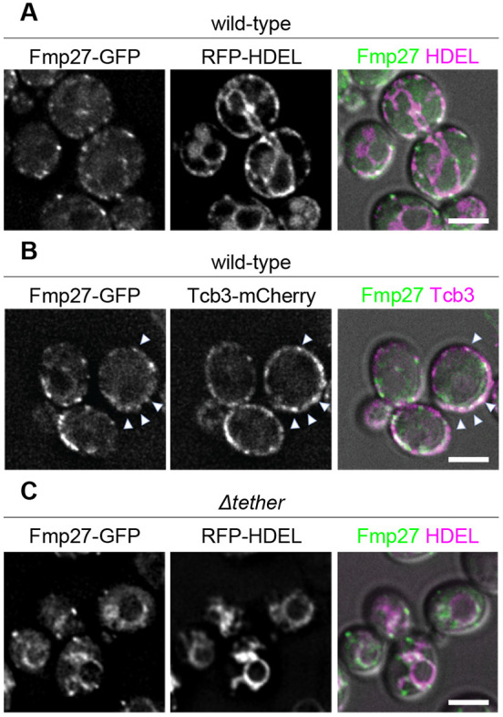
Fig. 1.

Fmp27 localizes to ER-PM contact sites. (A) Live-cell imaging of endogenously tagged Fmp27–GFP (green) shows that Fmp27 colocalizes with the pan-ER marker RFP–HDEL (magenta) and is enriched in puncta at the cell cortex. RFP–HDEL is expressed from a plasmid. (B) Live-cell imaging of Fmp27–GFP (green) with the ER–PM tether Tcb3–mCherry (magenta) confirms that Fmp27 is enriched at ER–PM contact sites (arrowheads highlight colocalization). (C) Live-cell imaging of endogenously tagged Fmp27–GFP (green) in the Δtether background with RFP-HDEL (magenta) shows that Fmp27 localizes to collapsed ER upon loss of ER-PM tethers. RFP–HDEL is expressed from a plasmid. Images shown are representative of three independent experiments with n≥3 images acquired per replicate. Scale bars: 2 µm.
